Left renal vein
The left renal vein drains the left kidney into the inferior vena cava. It is longer than the right renal vein, about 8.5 cm on average, and crosses in front of the aorta on the way. (StatPearls, Abdomen and Pelvis, Renal Veins; Gray’s Anatomy (1918), The Veins of the Abdomen and Pelvis)
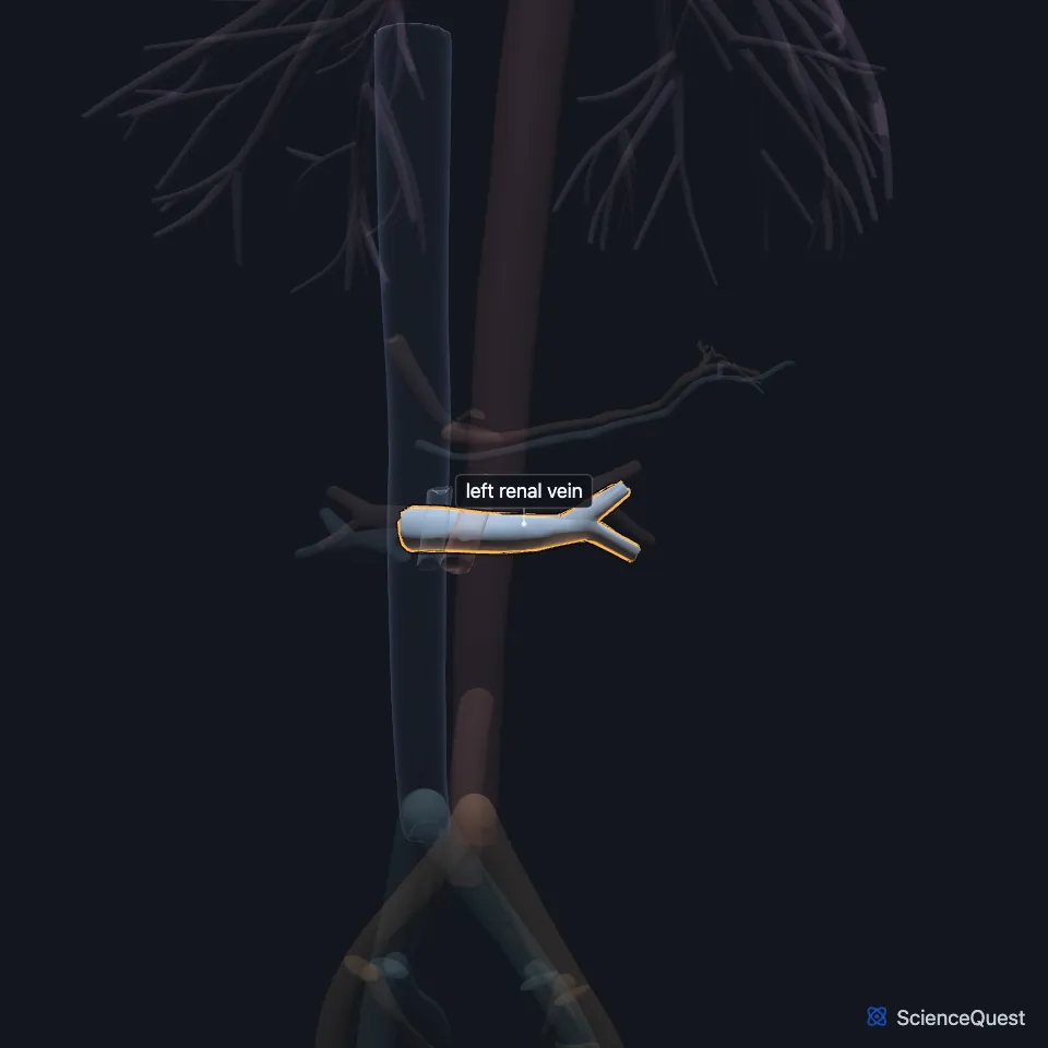
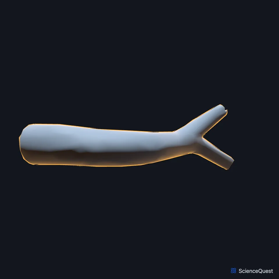
Where is the left renal vein?
It forms at the hilum of the kidney from the segmental veins and runs across to the medial side of the inferior vena cava, passing in front of the aorta and just below the superior mesenteric artery. (StatPearls, Abdomen and Pelvis, Renal Veins)
At the hilum the renal vein usually lies in front of and below the renal artery. Gray’s Anatomy (1918) notes that the left vein opens into the inferior vena cava a little higher than the right. (StatPearls, Abdomen and Pelvis, Renal Veins; Gray’s Anatomy (1918), The Veins of the Abdomen and Pelvis)
What does the left renal vein drain?
Besides the kidney, it receives the left adrenal vein and a branch of the inferior phrenic vein along its upper edge, and the left gonadal vein along its lower edge. (StatPearls, Abdomen and Pelvis, Renal Veins; Gray’s Anatomy (1918), The Veins of the Abdomen and Pelvis)
In about 75% of people, lumbar or hemiazygos branches also join its back surface. The right renal vein usually takes no tributaries outside the kidney. (StatPearls, Abdomen and Pelvis, Renal Veins)
What lies next to the left renal vein?
On its way to the inferior vena cava it passes between the superior mesenteric artery and the aorta. Fat behind the peritoneum and the third part of the duodenum keep a space open between the two arteries. (StatPearls, Abdomen and Pelvis, Renal Veins and Nutcracker Syndrome and Left Renal Vein Entrapment)
What is nutcracker syndrome?
Nutcracker syndrome is symptomatic compression of the left renal vein, often between the aorta and the superior mesenteric artery when the angle between them falls below 40 degrees. It can cause flank pain, blood or protein in the urine, and a varicocele. (StatPearls, Nutcracker Syndrome and Left Renal Vein Entrapment)
The same compression found on imaging without symptoms is called the nutcracker phenomenon and counts as a normal variant. (StatPearls, Nutcracker Syndrome and Left Renal Vein Entrapment)
Why are varicoceles more common on the left?
Varicoceles occur far more often on the left. The left testicular vein drains at an acute angle into the left renal vein, where pressure is relatively high, and this is thought to favor valve failure and backflow into the testicular vein. (StatPearls, Abdomen and Pelvis, Renal Veins)
How does the left renal vein vary?
A circumaortic left renal vein sends one branch in front of the aorta and one behind it. A retroaortic left renal vein, present in about 3% of people, passes only behind the aorta and can be squeezed against the spine, which is posterior nutcracker syndrome. (StatPearls, Abdomen and Pelvis, Renal Veins)
Why is the left kidney often taken for transplant?
Its renal vein is longer, so the left kidney is commonly suggested for transplant over the right. (StatPearls, Abdomen and Pelvis, Renal Veins)
Where the sources differ
StatPearls articles give different normal figures for the aorta and the superior mesenteric artery. The renal veins article gives a typical angle of 90 degrees and the nutcracker syndrome article a 4 to 5 mm space between the vessels; the superior mesenteric artery syndrome article gives an angle of about 38 to 65 degrees and a distance of 10 to 28 mm. (StatPearls, Abdomen and Pelvis, Renal Veins, Nutcracker Syndrome and Left Renal Vein Entrapment and Superior Mesenteric Artery Syndrome)
| Feature | Detail and source |
|---|---|
| Drains | Left kidney (StatPearls, Abdomen and Pelvis, Renal Veins) |
| Ends in | Inferior vena cava (StatPearls, Abdomen and Pelvis, Renal Veins) |
| Length | About 8.5 cm on average (StatPearls, Abdomen and Pelvis, Renal Veins) |
| Tributaries | Left adrenal vein, a branch of the inferior phrenic vein, left gonadal vein (StatPearls, Abdomen and Pelvis, Renal Veins) |
Touches in this model
Measured on the BodyParts3D meshes: surfaces within 2 mm of the left renal vein, nearest first.
- Descending aorta in contact
- Inferior vena cava in contact
- Left renal artery in contact
- Right renal artery in contact
- Superior mesenteric artery in contact
- Superior mesenteric vein in contact
- Left kidney in contact
- Left ureter in contact
- Pancreas 0.6 mm away
- Left psoas major 1.4 mm away
- Skin 1.5 mm away
Sources
Sources read between and
- StatPearls, Anatomy, Abdomen and Pelvis, Renal Veins. Cole S. Bowdino; Justin Owens; Palma M. Shaw. Last update . CC BY-NC-ND 4.0. Read .
- Anatomy of the Human Body (Gray’s Anatomy), 20th US edition, 1918, Angiology, The Veins of the Lower Extremity, Abdomen, and Pelvis: the external iliac, hypogastric and common iliac veins, and the renal veins among the tributaries of the inferior vena cava, pages 672 to 679; Internet Archive scan of the 1918 Lea and Febiger printing, terminology of 1918 (hypogastric for internal iliac, spermatic for testicular, hemorrhoidal for rectal). Henry Gray; edited by Warren H. Lewis. Public domain. Read .
- StatPearls, Nutcracker Syndrome and Left Renal Vein Entrapment. Dana Penfold; Stephen W. Leslie; Saran Lotfollahzadeh. Last update . CC BY-NC-ND 4.0. Read .
- StatPearls, Superior Mesenteric Artery Syndrome. Eric Goosenberg; Sara A. Collier. Last update . CC BY-NC-ND 4.0. Read .
The values are facts and free to use; this page’s selection and presentation are © 2026 ScienceQuest.