Internal iliac artery
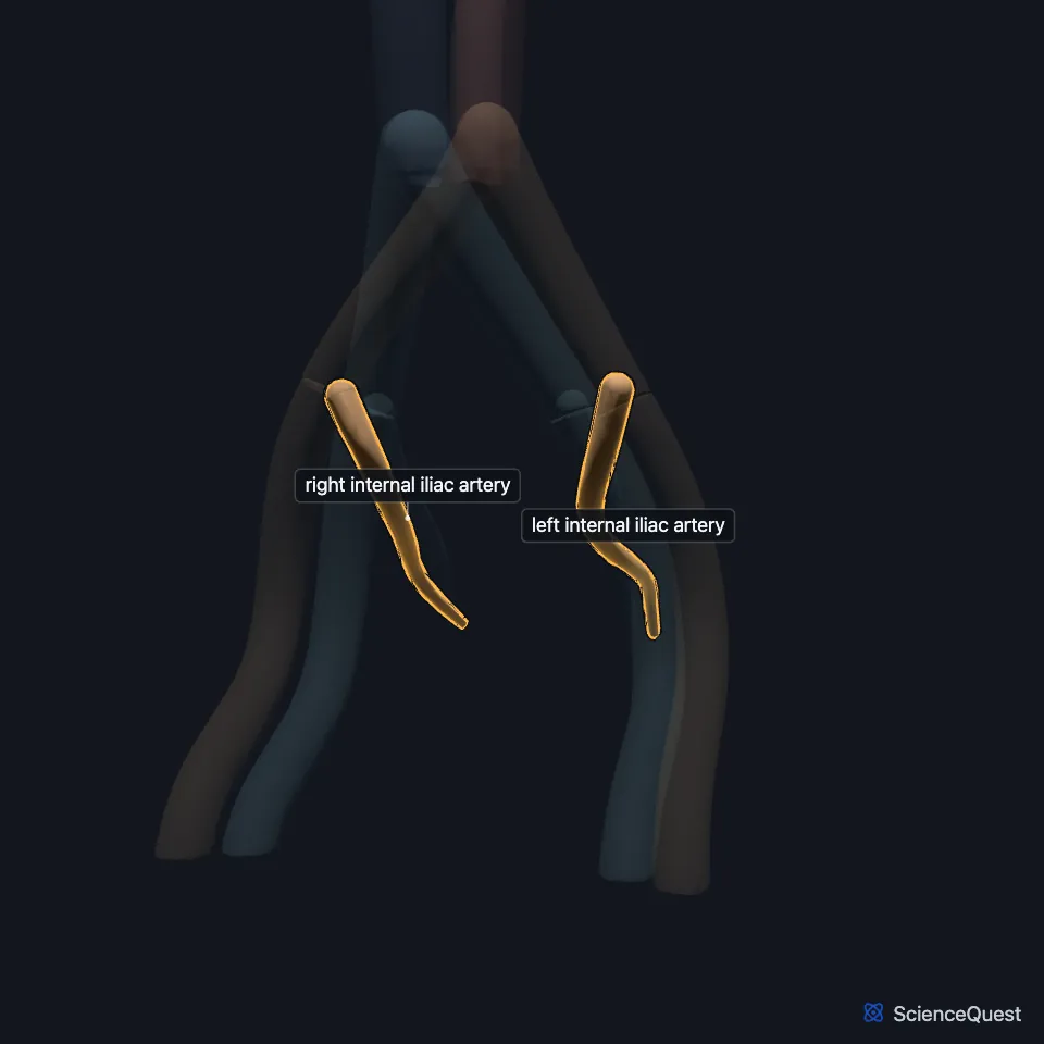
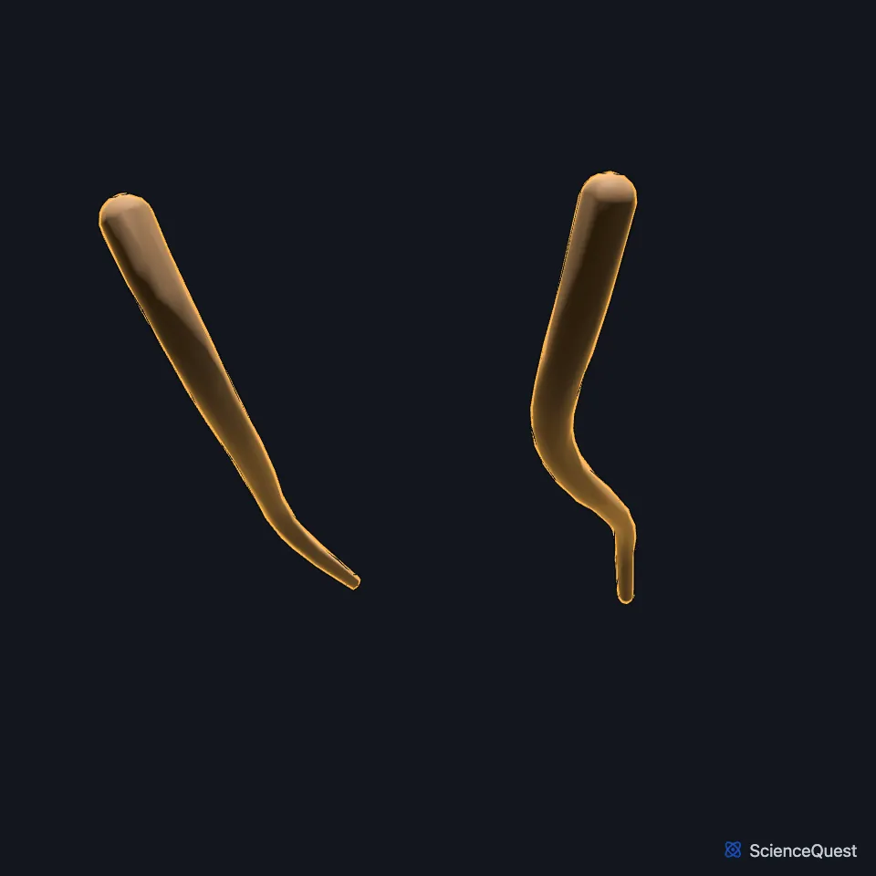
The internal iliac artery, also called the hypogastric artery, is the main artery of the pelvic organs. It also supplies the pelvic wall, the perineum, the buttock and the thigh. (StatPearls, Abdomen and Pelvis: Internal Iliac Arteries; Gray’s Anatomy (1918), The Common Iliac Arteries)
Where is the internal iliac artery?
It starts where the common iliac artery divides, usually in front of the sacroiliac joint, then crosses the pelvic brim into the lesser pelvis. (StatPearls, Abdomen and Pelvis: Internal Iliac Arteries)
Near the upper edge of the greater sciatic foramen it splits into anterior and posterior divisions. Gray’s Anatomy (1918) describes it as short and thick, about 4 cm long. (StatPearls, Abdomen and Pelvis: Internal Iliac Arteries; Gray’s Anatomy (1918), The Common Iliac Arteries)
What does the internal iliac artery supply?
Its visceral branches supply the bladder, rectum and urethra in both sexes, the prostate, ductus deferens and seminal vesicles in men, and the uterus and vagina in women. (StatPearls, Abdomen and Pelvis: Internal Iliac Arteries)
The internal pudendal artery is the main supply of the perineum, and the superior and inferior gluteal arteries supply the buttock muscles, among them the gluteus maximus and piriformis. (StatPearls, Abdomen and Pelvis: Internal Iliac Arteries)
What are the branches of the internal iliac artery?
The anterior division commonly gives 6 branches: the umbilical, obturator, inferior vesical (in men), middle rectal, internal pudendal and inferior gluteal arteries. (StatPearls, Abdomen and Pelvis: Internal Iliac Arteries)
The posterior division gives the iliolumbar, lateral sacral and superior gluteal arteries, the superior gluteal being the largest. (StatPearls, Abdomen and Pelvis: Internal Iliac Arteries)
The branching varies a great deal. The obturator artery comes from the external iliac artery, directly or through the inferior epigastric artery, in about 19% of people. (StatPearls, Abdomen and Pelvis: Internal Iliac Arteries)
What lies next to the internal iliac artery?
The ureter lies in front of it and the internal iliac vein behind and to its medial side. The external iliac vein, obturator nerve and obturator vein lie behind and to its lateral side, and the psoas major and obturator internus to its lateral side. (StatPearls, Abdomen and Pelvis: Internal Iliac Arteries)
What did the internal iliac artery do before birth?
Before birth it sends fetal blood to the placenta through the umbilical artery. Gray’s Anatomy (1918) notes that in the fetus it is twice the size of the external iliac artery and is the direct continuation of the common iliac. (StatPearls, Abdomen and Pelvis: Internal Iliac Arteries; Gray’s Anatomy (1918), The Common Iliac Arteries)
After birth the far part of the umbilical artery closes into a fibrous cord, while the near part stays open and gives the superior vesical artery to the bladder. (StatPearls, Abdomen and Pelvis: Internal Iliac Arteries; Gray’s Anatomy (1918), The Common Iliac Arteries)
That cord is the medial umbilical ligament, which runs up beside the bladder to the umbilicus. Gray’s Anatomy (1918) uses the older name lateral umbilical ligament, or obliterated hypogastric artery, for the same cord. (StatPearls, Abdomen and Pelvis: Internal Iliac Arteries; FMA, FMA14077; Gray’s Anatomy (1918), The Common Iliac Arteries)
| Feature | Detail and source |
|---|---|
| Arises from | Common iliac artery (StatPearls, Abdomen and Pelvis: Internal Iliac Arteries) |
| Divides into | Anterior and posterior divisions (StatPearls, Abdomen and Pelvis: Internal Iliac Arteries) |
| Also called | Hypogastric artery (StatPearls, Abdomen and Pelvis: Internal Iliac Arteries; Gray’s Anatomy (1918), The Common Iliac Arteries) |
Touches in this model
Measured on the BodyParts3D meshes: surfaces within 2 mm of the internal iliac artery, nearest first, 12 of 13.
- Left common iliac artery in contact
- Left external iliac artery in contact
- Left external iliac vein in contact
- Right common iliac artery in contact
- Right external iliac artery in contact
- Right internal iliac vein in contact
- Colon in contact
- Left piriformis in contact
- Sacrum in contact
- Skin in contact
- Right external iliac vein 1.1 mm away
- Left internal iliac vein 1.2 mm away
Sources
Sources read between and
- StatPearls, Anatomy, Abdomen and Pelvis: Internal Iliac Arteries. Nicolas Zaunbrecher; Tafline C. Arbor; Navdeep S. Samra. Last update . CC BY-NC-ND 4.0. Read .
- Anatomy of the Human Body (Gray’s Anatomy), 20th US edition, 1918, Angiology, The Common Iliac Arteries (Aa. Iliacae Communes); Internet Archive scan of the 1918 Lea and Febiger printing, terminology of 1918. Henry Gray; edited by Warren H. Lewis. Public domain. Read .
- Foundational Model of Anatomy, via the EMBL-EBI Ontology Lookup Service, Medial umbilical ligament (FMA14077), preferred name and definition. Not printed on the page. Read .
The values are facts and free to use; this page’s selection and presentation are © 2026 ScienceQuest.