Pyramidalis
The pyramidalis is a small triangular muscle low in the front of the abdomen, in front of the rectus abdominis and inside its sheath. Some people lack it on one or both sides. (Gray 1918, 6d. Muscles of the Abdomen; StatPearls, Rectus Sheath)
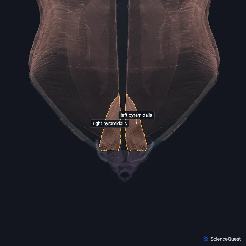
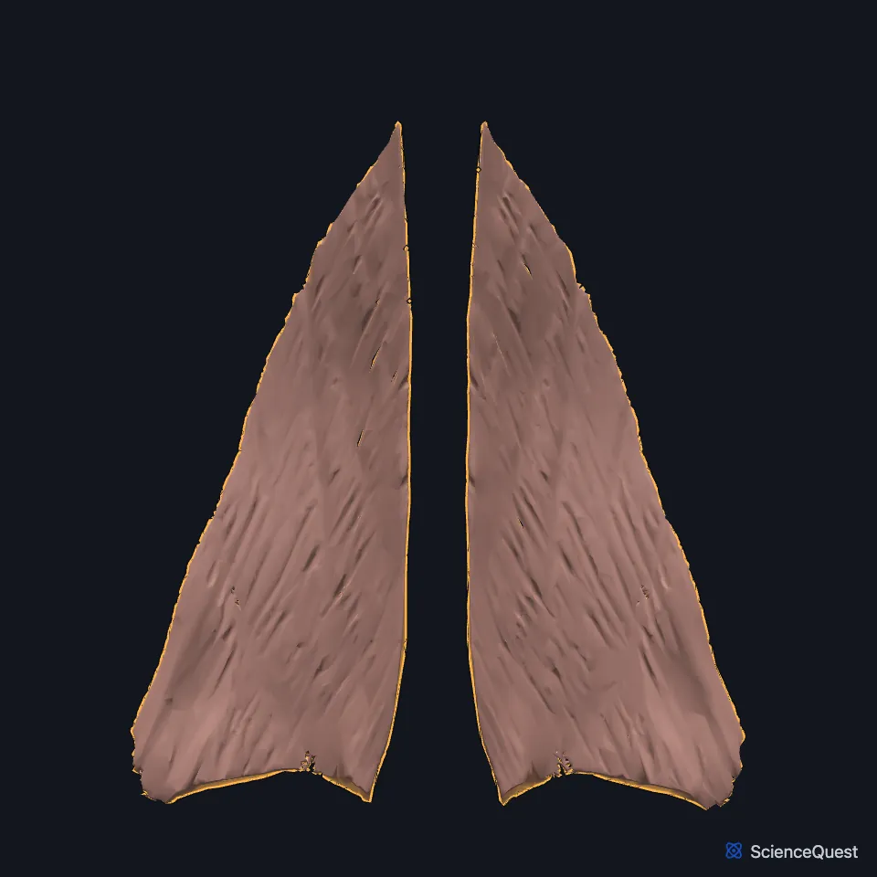
Open it in the Abdominal Muscles Explorer See it alone in 3D
Where is the pyramidalis?
It arises by tendinous fibers from the front of the pubis and the anterior pubic ligament. It narrows as it rises and ends in a point on the linea alba, midway between the umbilicus and the pubis. (Gray 1918, 6d. Muscles of the Abdomen)
A thin fascial sheath separates it from the lower fibers of the rectus abdominis. The authors of a 2024 imaging study list the pubic crest, the anterior pubic ligament and the inguinal ligament as its origins. (Short et al. 2024, Surg Radiol Anat)
The rectus sheath holds the rectus abdominis and the pyramidalis. Below the arcuate line, only the transversalis fascia covers the back of both muscles. (StatPearls, Rectus Sheath)
What does the pyramidalis do?
It tenses the linea alba when it contracts. (StatPearls, Abdominal Wall and Anterolateral Abdominal Wall Fascia)
In a 2026 ultrasound study, electrical stimulation of the pyramidalis in 10 people made the linea alba stiffer. Its authors read this as support for a role that had gone unvalidated until then. (Short et al. 2026, J Anat)
StatPearls adds that the rectus abdominis and pyramidalis together flex the lumbar spine. (StatPearls, Rectus Sheath)
What supplies the pyramidalis?
The subcostal nerve, which the anterior ramus of T12 forms, supplies it. Gray’s 1918 text names the twelfth thoracic nerve. (StatPearls, Rectus Sheath and Thorax, Muscles; Gray 1918, 6d. Muscles of the Abdomen)
In a CT angiography study of 32 women, a dedicated muscular branch of the inferior epigastric artery supplied 68% of the muscles studied. (Short et al. 2024, Surg Radiol Anat)
How often is the pyramidalis missing?
StatPearls reports it absent in about 1 person in 5. Gray’s 1918 text adds that the lower rectus abdominis grows larger when it is missing, that it can be double on one side, and that the two sides can differ in size. (StatPearls, Rectus Sheath; Gray 1918, 6d. Muscles of the Abdomen)
Where the sources differ
StatPearls gives it as absent in 20% of people, or present on both sides in 80%. Two imaging studies from one Adelaide group found it on both sides in 75%, on one side only in 6% to 10%, and absent in 15% to 19%. (StatPearls, Rectus Sheath and Abdominal Wall; Short et al. 2024, Surg Radiol Anat; Short et al. 2026, J Anat)
| Feature | Detail and source |
|---|---|
| Origin | Front of the pubis and the anterior pubic ligament (Gray 1918, 6d. Muscles of the Abdomen) |
| Insertion | Linea alba, midway between the umbilicus and the pubis (Gray 1918, 6d. Muscles of the Abdomen) |
| Action | Tenses the linea alba (StatPearls, Abdominal Wall) |
| Nerve | Subcostal nerve (T12) (StatPearls, Rectus Sheath; Gray 1918, 6d. Muscles of the Abdomen) |
| Absent | In about 1 person in 5 (StatPearls); 15% to 19% in two imaging studies (StatPearls, Rectus Sheath; Short et al. 2024, Surg Radiol Anat; Short et al. 2026, J Anat) |
Touches in this model
Measured on the BodyParts3D meshes: surfaces within 2 mm of the pyramidalis, nearest first, 12 of 15.
- Left inguinal ligament in contact
- Left internal oblique in contact
- Left rectus abdominis in contact
- Left transversus abdominis in contact
- Linea alba in contact
- Right inguinal ligament in contact
- Right internal oblique in contact
- Right rectus abdominis in contact
- Right transversus abdominis in contact
- Left hip bone in contact
- Left pectineus in contact
- Right hip bone in contact
Sources
Sources read
- Gray’s Anatomy of the Human Body, 20th US edition (1918), IV. Myology, 6d. The Muscles and Fasciae of the Abdomen. Henry Gray. Public domain. Read .
- StatPearls, Anatomy, Abdomen and Pelvis, Rectus Sheath. Karlin E. Sevensma; Logan Leavitt; Kerent D. Pihl. Last update . CC BY-NC-ND 4.0. Read .
- Surgical and Radiologic Anatomy, Morphology and arterial supply of the pyramidalis muscle in an Australian female population using computed tomography angiography, Surg Radiol Anat 46, from page 1865 (full text, PMC11458779, doi 10.1007/s00276-024-03471-1). Short CL, Crotti TN, Algate K, Gladman MA, Barras CD. CC BY 4.0. Read .
- StatPearls, Anatomy, Abdomen and Pelvis: Abdominal Wall. William Flynn; Paula Vickerton. Last update . CC BY-NC-ND 4.0. Read .
- StatPearls, Anatomy, Abdomen and Pelvis: Anterolateral Abdominal Wall Fascia. Leandra A. Jelinek; Stephanie Scharbach; Sarang Kashyap; Troy Ferguson. Last update . CC BY-NC-ND 4.0. Read .
- Journal of Anatomy, An investigation of the functional significance of the pyramidalis muscle using shear wave elastography, J Anat 249, from page 1130 (full text, PMC13399048, doi 10.1111/joa.70160). Short CL, Morrison T, Crotti TN, Algate K, Gladman MA, Barras CD. CC BY-NC-ND 4.0. Read .
- StatPearls, Anatomy, Thorax, Muscles. Alex Tang; Bruno Bordoni. Last update . CC BY-NC-ND 4.0. Read .
The values are facts and free to use; this page’s selection and presentation are © 2026 ScienceQuest.