Adductor longus
The adductor longus is a muscle of the medial compartment of the thigh. It runs from the body of the pubis to the middle third of the femur and adducts the thigh at the hip. (StatPearls, Medial Thigh Muscles; OpenStax A&P 2e, 11.6; Gray’s Anatomy (1918), Medial Femoral Muscles)
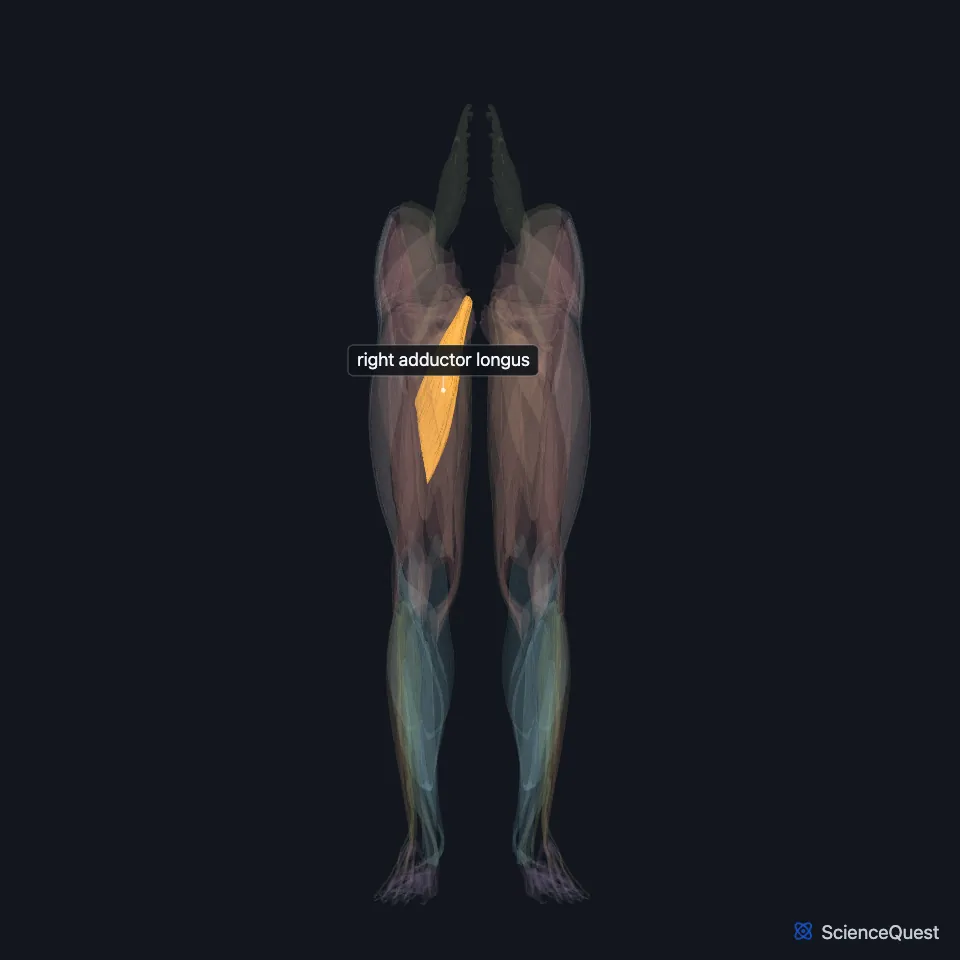
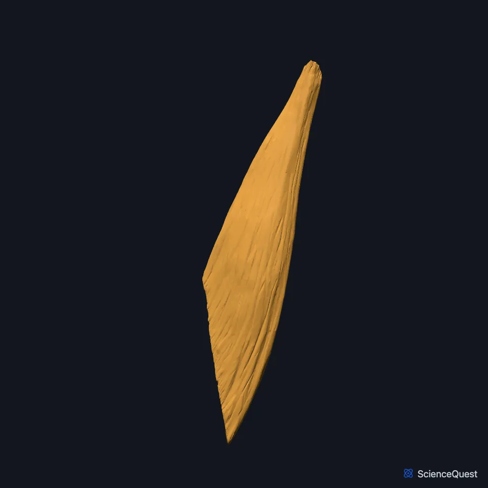
Open it in the Lower Limb Muscles Explorer See it alone in 3D
Where is the adductor longus?
It shares the medial compartment with adductor brevis, adductor magnus, pectineus and gracilis. (OpenStax A&P 2e, 11.6; Gray’s Anatomy (1918), Medial Femoral Muscles)
It forms the medial side of the femoral triangle, with sartorius laterally and the inguinal ligament above. (StatPearls, Thigh Femoral Nerve)
Lower down it forms the upper part of the floor of the adductor canal, with adductor magnus below it. (StatPearls, Anterior Thigh Muscles)
Where does the adductor longus attach?
About 40% of the muscle arises by tendon fibers near the pubic tubercle, at the front, and 60% by muscle fibers from the back of the pubic symphysis. (StatPearls, Medial Thigh Muscles)
It ends as a flat tendon on the linea aspera, in the middle third of the shaft of the femur. (StatPearls, Thigh Muscles and Medial Thigh Muscles)
Longus is Latin for long, and brevis, in its neighbor adductor brevis, for short. (OpenStax A&P 2e, 11.2; Lewis and Short, longus and brevis; Gray’s Anatomy (1918), Medial Femoral Muscles)
What does the adductor longus do?
It pulls the thigh toward the midline and also helps flex it at the hip. (OpenStax A&P 2e, 11.6; Gray’s Anatomy (1918), Medial Femoral Muscles)
What supplies the adductor longus?
The anterior division of the obturator nerve supplies it, from L2, L3 and L4. That branch of the nerve runs between adductor longus and adductor brevis. (StatPearls, Thigh Muscles and Obturator Nerve)
The posterior branch of the obturator nerve occasionally supplies it as well. (StatPearls, Obturator Nerve)
It shares its blood supply with adductor brevis: the obturator and medial circumflex femoral arteries. (StatPearls, Thigh Muscles)
Where the sources differ
Sources differ on which edge of the muscle bounds the femoral triangle. The StatPearls anterior thigh article gives the lateral border of adductor longus; the adductor canal article gives its medial border, and notes that some sources put the triangle’s apex at its lateral border. (StatPearls, Anterior Thigh Muscles and Adductor Canal)
OpenStax says the adductors longus, brevis and magnus can rotate the thigh either way, by the placement of the foot. The StatPearls thigh muscles article says longus and brevis rotate the hip laterally, and Gray’s Anatomy (1918) says the slant of the adductors' attachments makes them turn the thigh outward. (OpenStax A&P 2e, 11.6; StatPearls, Thigh Muscles; Gray’s Anatomy (1918), Medial Femoral Muscles)
| Feature | Detail and source |
|---|---|
| Origin | Body of the pubis and the front lower pubic ramus (StatPearls, Medial Thigh Muscles) |
| Insertion | Linea aspera, middle third of the femoral shaft (StatPearls, Medial Thigh Muscles) |
| Action | Adducts and flexes the thigh at the hip (OpenStax A&P 2e, 11.6; Gray’s Anatomy (1918), Medial Femoral Muscles) |
| Nerve | Obturator nerve, anterior division (L2 to L4) (StatPearls, Thigh Muscles) |
Touches in this model
Measured on the BodyParts3D meshes: surfaces within 2 mm of the adductor longus, nearest first, 12 of 13.
- Right adductor brevis in contact
- Right adductor magnus in contact
- Right gracilis in contact
- Right pectineus in contact
- Right sartorius in contact
- Right vastus medialis in contact
- Right deferent duct in contact
- Right femur in contact
- Right hip bone in contact
- Right inguinal ligament in contact
- Skin in contact
- Corpus cavernosum of penis 1.0 mm away
Sources
- StatPearls, Anatomy, Bony Pelvis and Lower Limb: Medial Thigh Muscles. Jordan L. Ramage; Matthew A. Varacallo. Last update August 21, 2023. CC BY-NC-ND 4.0. Read 2026-10-25.
- OpenStax Anatomy and Physiology 2e, section 11.6, Appendicular Muscles of the Pelvic Girdle and Lower Limbs. CC BY-NC-SA 4.0. Read 2026-10-25.
- Anatomy of the Human Body (Gray’s Anatomy), 20th US edition, 1918, Myology, The Muscles and Fasciae of the Thigh, 2. The Medial Femoral Muscles, pages 471 to 474; Internet Archive scan of the 1918 Lea and Febiger printing, terminology of 1918. Henry Gray; edited by Warren H. Lewis. Public domain. Read 2026-10-25.
- StatPearls, Anatomy, Bony Pelvis and Lower Limb: Thigh Femoral Nerve. Nader A. Refai; Asa C. Black; Prasanna Tadi. Last update August 22, 2023. CC BY-NC-ND 4.0. Read 2026-10-25.
- StatPearls, Anatomy, Bony Pelvis and Lower Limb: Anterior Thigh Muscles. Ayesha Khan; Abdul Arain. Last update March 17, 2023. CC BY-NC-ND 4.0. Read 2026-10-25.
- StatPearls, Anatomy, Bony Pelvis and Lower Limb: Thigh Muscles. Basem Attum; Matthew A. Varacallo. Last update December 17, 2022. CC BY-NC-ND 4.0. Read 2026-10-25.
- OpenStax Anatomy and Physiology 2e, section 11.2, Naming Skeletal Muscles. CC BY-NC-SA 4.0. Read 2026-10-25.
- A Latin Dictionary (Lewis and Short, 1879), entry longus, read in the Lewis and Short text served by Logeion, University of Chicago. Charlton T. Lewis; Charles Short. Public domain. Read 2026-10-25.
- A Latin Dictionary (Lewis and Short, 1879), entry brevis, read in the Lewis and Short text served by Logeion, University of Chicago. Charlton T. Lewis; Charles Short. Public domain. Read 2026-10-25.
- StatPearls, Anatomy, Abdomen and Pelvis, Obturator Nerve. Matthew Koh; Brian Markovich. Last update July 24, 2023. CC BY-NC-ND 4.0. Read 2026-10-25.
- StatPearls, Anatomy, Abdomen and Pelvis: Adductor Canal (Subsartorial Canal, Hunter Canal). Allan Migirov; Tafline C. Arbor; Renato C. Vilella. Last update January 9, 2024. CC BY-NC-ND 4.0. Read 2026-10-25.
The values are facts and free to use; this page’s selection and presentation are © 2026 ScienceQuest.