External anal sphincter
The external anal sphincter is a tube of striated muscle around the anal canal, under voluntary control. Its tone keeps the anal opening closed, and it relaxes for defecation. (StatPearls, Perineal Body and Anal Canal)
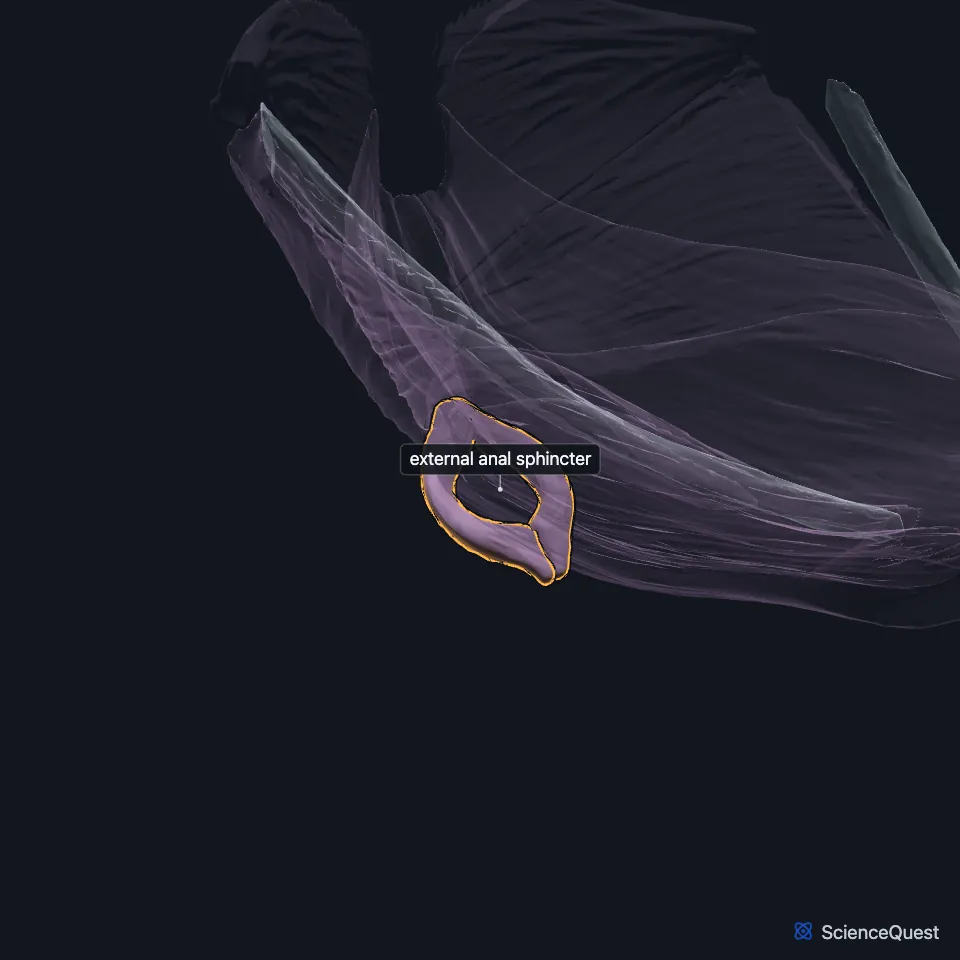
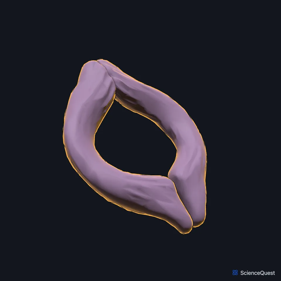
Open it in the Abdominal Muscles Explorer See it alone in 3D
Where is the external anal sphincter?
It encircles the anal canal as a sleeve about 3 mm thick, about 4 cm long in men and 3 cm in women. Its right and left halves work as one. (StatPearls, Perineal Body)
In front it attaches to the perineal body, the central tendon of the perineum. Gray’s 1918 text counts six muscles meeting there, this sphincter among them. (StatPearls, Perineal Body; Gray 1918, Muscles of the Perineum)
What are the parts of the external anal sphincter?
The classic description gives it three parts. The deep part is a thick ring around the top of the internal sphincter, its fibers fused with the puborectalis. (StatPearls, Perineal Body)
The superficial part runs from the perineal body in front to the last coccygeal segment behind, through the anococcygeal raphe. (StatPearls, Perineal Body)
The subcutaneous part is a flat band about 15 mm wide, just under the skin at the anal opening. (StatPearls, Perineal Body)
What does the external anal sphincter do?
When the rectum fills, a reflex relaxes the internal sphincter, and the external sphincter holds stool in the anal canal until the person is ready to defecate. (StatPearls, Anal Canal)
Gray’s 1918 text describes it as always in tonic contraction, with no muscle opposing it, and able to tighten at will to close the anus harder. (Gray 1918, Muscles of the Perineum)
Babies are born without full voluntary control of it, and most children gain that control at 18 to 24 months. (StatPearls, Anal Canal)
What lies inside the external anal sphincter, and what supplies it?
The internal anal sphincter, an involuntary muscle, lies inside it, and the conjoint longitudinal muscle coat runs down between the two. (StatPearls, Anal Canal and Perineal Body)
The inferior rectal nerve, the first branch of the pudendal nerve, carries motor fibers to the sphincter, and this supply gives voluntary control of defecation. Muscular branches of the perineal nerve, a terminal branch of the pudendal nerve, also reach its front part. (StatPearls, Pudendal Nerve and Perineal Body)
Gray’s 1918 text names a branch of the fourth sacral nerve and twigs from the pudendal nerve’s inferior hemorrhoidal branch. The Foundational Model of Anatomy uses the name inferior rectal nerve and lists inferior hemorrhoidal nerve as a synonym. (Gray 1918, Muscles of the Perineum; FMA, Inferior rectal nerve)
Where the sources differ
StatPearls gives the classical three parts and notes studies that see loops varying between people where others see one continuous ring. Gray’s 1918 text describes two layers, superficial and deep. (StatPearls, Perineal Body; Gray 1918, Muscles of the Perineum)
| Feature | Detail and source |
|---|---|
| Parts | Deep, superficial and subcutaneous, in the classic account (StatPearls, Perineal Body) |
| Attachments | Perineal body in front; anococcygeal raphe and the last coccygeal segment behind (StatPearls, Perineal Body) |
| Action | Keeps the anal opening closed; relaxes for defecation (StatPearls, Anal Canal) |
| Control | Voluntary (StatPearls, Anal Canal) |
| Nerve | Inferior rectal nerve, from the pudendal nerve; front part also from the perineal nerve (StatPearls, Pudendal Nerve and Perineal Body; Gray 1918, Muscles of the Perineum; FMA, Inferior rectal nerve) |
Touches in this model
Measured on the BodyParts3D meshes: surfaces within 2 mm of the external anal sphincter, nearest first.
- Left puborectalis in contact
- Right puborectalis in contact
- Rectum in contact
- Skin in contact
Sources
Sources read between and
- StatPearls, Anatomy, Abdomen and Pelvis, Perineal Body. Bruno Bordoni; Marjorie V. Launico. Last update . CC BY-NC-ND 4.0. Read .
- StatPearls, Anatomy, Abdomen and Pelvis: Anal Canal. Abdelwahab Ahmed; Tafline C. Arbor; Waqar A. Qureshi. Last update . CC BY-NC-ND 4.0. Read .
- Gray’s Anatomy of the Human Body, 20th US edition (1918), IV. Myology, The Muscles and Fasciae of the Perineum (section numbered 1F on the Bartleby page). Henry Gray. Public domain. Read .
- StatPearls, Anatomy, Abdomen and Pelvis, Pudendal Nerve. Kevin J. Kinter; Bruce W. Newton. Last update . CC BY-NC-ND 4.0. Read .
- Foundational Model of Anatomy, FMA21865, Inferior rectal nerve (read on EBI OLS). FMA release terms, University of Washington. Read .
The values are facts and free to use; this page’s selection and presentation are © 2026 ScienceQuest.