Thorax Explorer
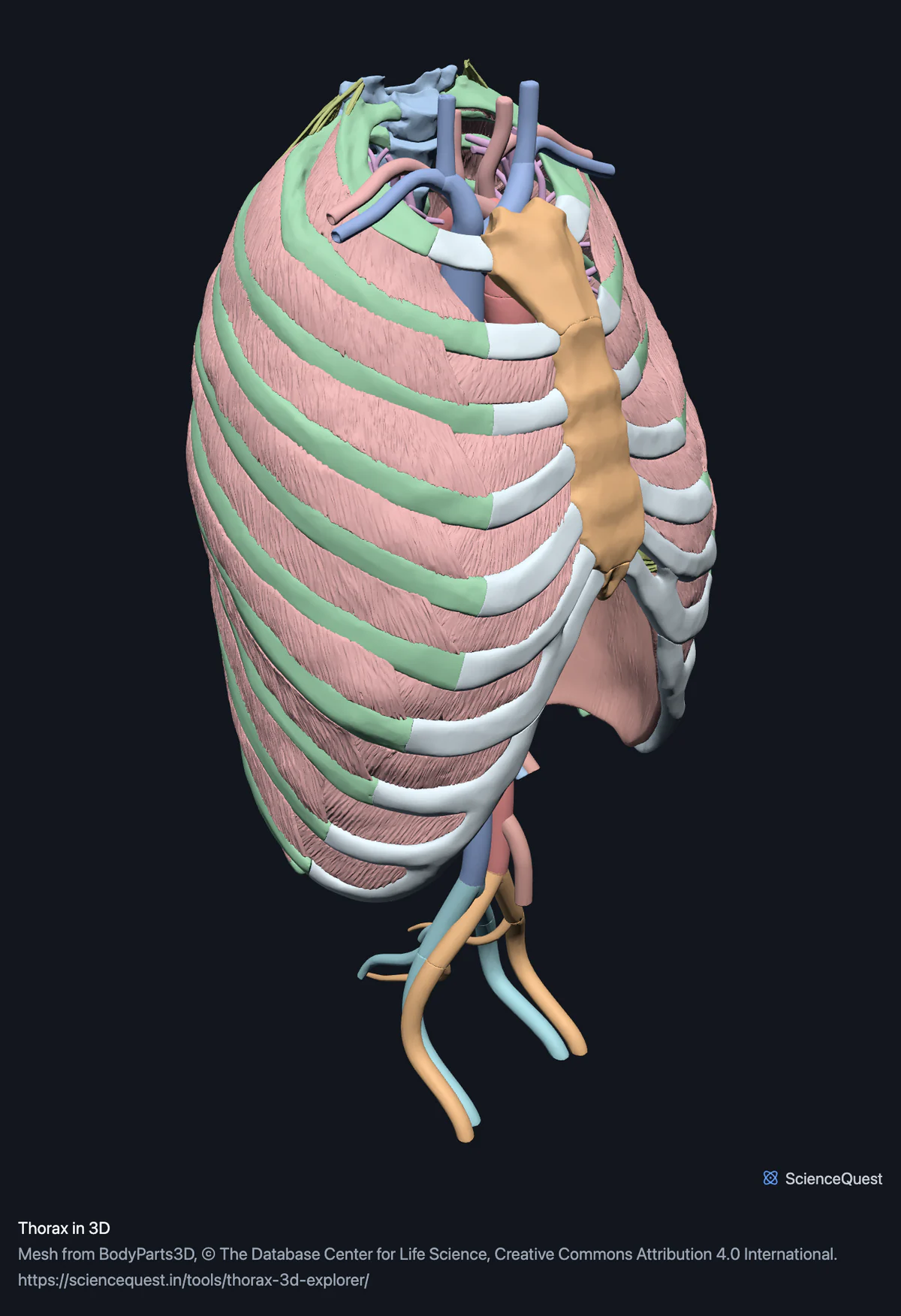
Rib cage, thoracic spine, heart, great vessels and chest wall muscles in one 3D scene: 130 structures, with the cage glassed to see through it.
Visualiser
Rib cage and thoracic spine, 39 bones in 55 meshes
Checking this device for a stored copy.
Start here
- Glass the Ribs band, then turn the model Glass keeps all 24 ribs visible and stops them taking clicks, so you can select the aorta through the cage. That is the mediastinum seen from outside its wall.
- Select the Coronary arteries band and save a figure Nine vessels labelled on the heart wall, with the dataset credit and licence burned into the image.
- Filter to Cartilage The 16 costal cartilage meshes on their own: the first to seventh on each side, and one more per side that the dataset names without a number. They are the reason the rib cage count and the bone count differ, and they are the part a dry skeleton never shows you.
Bone, heart, vessels and muscle. No lungs, because they are segmented with the abdominal organs and this page does not load that model. No nerves either, at any level: the only nerves the dataset publishes anywhere are the optic nerves, with their chiasm and tracts. In the other direction the page carries more than its name, because the vessel model is the whole trunk and 24 of its 42 structures are abdominal or pelvic.
Where this comes from
Segmented from imaging of one adult male body, not drawn by an artist, so the shapes are measurements. Two consequences worth knowing: it is one person’s anatomy rather than an average, and faint stepping on some surfaces is the resolution of the original scan rather than a rendering fault.
Meshes from BodyParts3D, © The Database Center for Life Science, under Creative Commons Attribution 4.0 International. Names and identifiers are the dataset’s own: Foundational Model of Anatomy identifiers and English labels, as its parts list gives them. Converted meshes are shared under the same licence.
Changes made: Meshes were converted from binary STL to an indexed mesh with 16-bit quantised vertex positions, and surface normals were recomputed from the geometry. No vertex was moved other than by that quantisation, whose worst-case error on any one coordinate is reported on the page. On this page it is at most 0.00432 mm, the largest among the 4 models the page can load.
Latin terms for 53 of the 130 structures in these models come from English Wikipedia anatomy infoboxes, under Creative Commons Attribution-ShareAlike 4.0, joined by Wikidata property P1402, Foundational Model of Anatomy identifier. Only a term a source states for that structure is used, never one constructed from a family name, which is why the rest carry no Latin rather than a guess.
Citing this tool
Last updated . Add the date you accessed it as well, which a citation of a page that can change asks for. If a specific view matters, cite the page address as it stands in your browser instead: it records what is selected and shown.
Why there are abdominal vessels on a page about the chest
Because the vessel model is loaded whole rather than cut down to the chest. The renal, coeliac, mesenteric and splenic vessels and the iliacs all arrive with the aorta, so 24 of the 42 vessel structures here sit below the diaphragm. That follows from how these pages are built: each one composes assets that have already been checked, mesh by mesh and axis by axis, rather than re-cutting them into new binaries with new digests and new chances to be wrong. The cost is a boundary that follows the segmentation rather than the region, and the honest way to pay it is to say so rather than to trim the scene quietly and leave a line in it that is not in the body.
Common questions
What does glassing the rib cage do?
It leaves the cage drawn but makes it translucent and unclickable, so you can see and select the aorta behind it. That combination is the point: hiding the ribs gives you a clear view of a mediastinum with no reference for where it sits, and leaving them solid gives you the reference and nothing to look at. Glass gives both, and because the ribs stop taking clicks you can pick a vessel through them without first switching anything off.
Why are there abdominal vessels on a page about the chest?
Because the vessel model is the whole trunk and this page loads it whole. Of its 42 structures, 24 are abdominal or pelvic: the renal, coeliac, mesenteric and splenic vessels and the iliacs. They are here because composing a region from assets that are already verified is worth more than a tidy boundary, and cutting them out would mean a new binary and a new digest for the sake of appearances. The alternative, hiding them quietly, would be worse: it would draw a line in the scene that is not in the body.
Where are the lungs?
In the chest and abdominal organs model, which this page does not load. That model is a single asset covering the trunk, so loading it to get the lungs would also bring the entire digestive tract into a page about the mediastinum. The regional abdomen page loads it instead, and the lungs arrive there as a consequence. If you want them beside the heart, the full body page’s Organs preset loads the organs, heart and vessels together over the skeleton.
Are the nerves of the chest wall included?
No, and not because they were left out. The only nerves BodyParts3D publishes anywhere are the optic nerves, with their chiasm and tracts. There are no intercostal nerves, no sympathetic chain and no phrenic nerve in the dataset to load, at any level below the neck. That is a limit of the source segmentation rather than a choice made here, which is why it is written on the page rather than discovered by looking for something that is not there.