Back Muscles Explorer
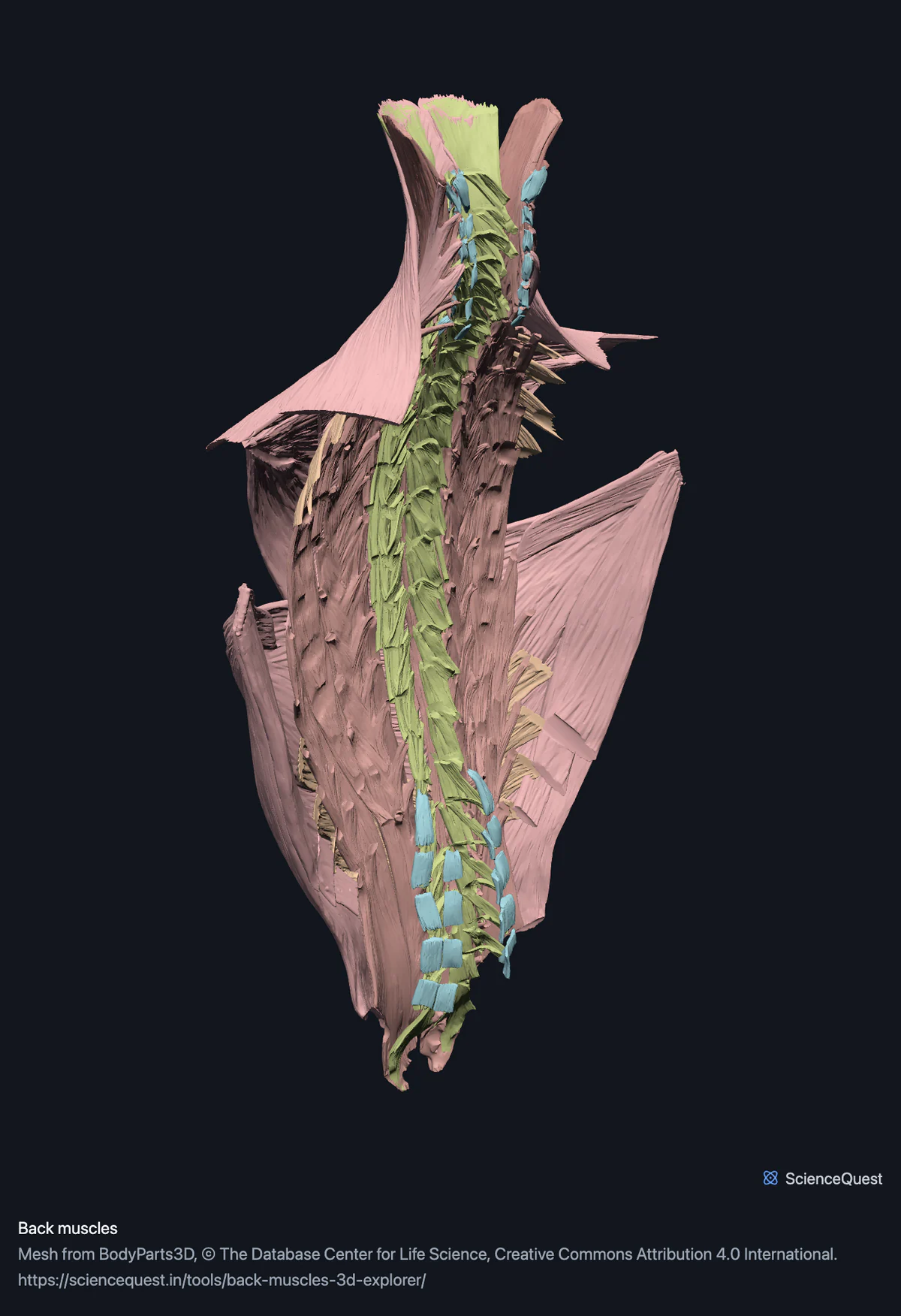
Turn 58 muscle meshes of the back in 3D through the six layers they are dissected in, from trapezius down to the rotatores and interspinales.
Visualiser
Back muscles, 58 structures
Checking this device for a stored copy.
Start here
- Hide the bands top down, in order Superficial, then intermediate, then splenius, then erector spinae. That is the order a back is dissected in, and each layer genuinely hides everything beneath it.
- Click Erector spinae, then Back Three columns, not one mass. Iliocostalis laterally, longissimus in the middle, spinalis medially. Here iliocostalis comes in cervicis, thoracis and lumborum parts, longissimus in capitis, cervicis and thoracis, and spinalis in cervicis and thoracis only.
- Show only the Segmental band The interspinales and intertransversarii, short muscles that join one spinous or transverse process to the next, published as sets rather than one per level. None of them can be seen from outside. The rotatores lie just as deep, but they are in the Transversospinal band.
Muscle only. No vertebrae, no ribs and no thoracolumbar fascia, which is a real absence for this region: the fascia is what latissimus dorsi and the erector spinae actually attach through. The deep muscles cannot be taken apart segment by segment.
- 58 structures
- 2795k triangles
- 14000 KB, on request
- 0.00511 mm worst error per axis
- 14 superficial layer
- 4 intermediate layer
- 4 splenius
- 16 erector spinae
- 13 transversospinal
- 7 segmental
Where this comes from
Segmented from imaging of one adult male body, not drawn by an artist, so the shapes are measurements. Two consequences worth knowing: it is one person’s anatomy rather than an average, and faint stepping on some surfaces is the resolution of the original scan rather than a rendering fault.
Meshes from BodyParts3D, © The Database Center for Life Science, under Creative Commons Attribution 4.0 International. Names and identifiers are the dataset’s own: Foundational Model of Anatomy identifiers and English labels, as its parts list gives them. Converted meshes are shared under the same licence.
Changes made: Meshes were converted from binary STL to an indexed mesh with 16-bit quantised vertex positions, and surface normals were recomputed from the geometry. No vertex was moved other than by that quantisation, whose worst-case error on any one coordinate is reported on the page.
Latin terms for 18 of the 58 structures come from English Wikipedia anatomy infoboxes, under Creative Commons Attribution-ShareAlike 4.0, joined by Wikidata property P1402, Foundational Model of Anatomy identifier. Only a term a source states for that structure is used, never one constructed from a family name, which is why the rest carry no Latin rather than a guess.
All 58 structures
Every structure the model contains, group by group and alphabetically within each. Each name opens the viewer with that structure selected, so a link can point at one structure rather than at the whole region.
Superficial layer 14
- ascending part of left trapezius muscle
- ascending part of right trapezius muscle
- descending part of left trapezius muscle
- descending part of right trapezius muscle
- left latissimus dorsi musculus latissimus dorsi muscle
- left levator scapulae musculus levator scapulae muscle
- left rhomboid major musculus rhomboideus major muscle
- left rhomboid minor musculus rhomboideus minor muscle
- right latissimus dorsi musculus latissimus dorsi muscle
- right levator scapulae musculus levator scapulae muscle
- right rhomboid major musculus rhomboideus major muscle
- right rhomboid minor musculus rhomboideus minor muscle
- transverse part of left trapezius muscle
- transverse part of right trapezius muscle
Intermediate layer 4
- left serratus posterior inferior musculus serratus posterior inferior muscle
- left serratus posterior superior musculus serratus posterior superior muscle
- right serratus posterior inferior musculus serratus posterior inferior muscle
- right serratus posterior superior musculus serratus posterior superior muscle
Splenius 4
- left splenius capitis musculus splenius capitis muscle
- left splenius cervicis musculus splenius cervicis muscle
- right splenius capitis musculus splenius capitis muscle
- right splenius cervicis musculus splenius cervicis muscle
Erector spinae 16
- left iliocostalis cervicis muscle
- left iliocostalis lumborum muscle
- left iliocostalis thoracis muscle
- left longissimus capitis muscle
- left longissimus cervicis muscle
- left longissimus thoracis muscle
- left spinalis cervicis muscle
- left spinalis thoracis muscle
- right iliocostalis cervicis muscle
- right iliocostalis lumborum muscle
- right iliocostalis thoracis muscle
- right longissimus capitis muscle
- right longissimus cervicis muscle
- right longissimus thoracis muscle
- right spinalis cervicis muscle
- right spinalis thoracis muscle
Transversospinal 13
- left cervical rotator muscle
- left lumbar rotator muscle
- left multifidus musculus multifidus muscle
- left semispinalis capitis muscle
- left semispinalis cervicis muscle
- left semispinalis thoracis muscle
- right cervical rotator muscle
- right lumbar rotator muscle
- right multifidus musculus multifidus muscle
- right semispinalis capitis muscle
- right semispinalis cervicis muscle
- right semispinalis thoracis muscle
- thoracic rotator muscle
Segmental 7
- set of anterior cervical intertransversarii muscle
- set of interspinales cervicis muscle
- set of interspinales lumborum muscle
- set of interspinales thoracis muscle
- set of lateral lumbar intertransversarius muscles muscle
- set of medial lumbar intertransversarius muscles muscle
- set of posterior cervical intertransversarii muscle
Citing this tool
Last updated . Add the date you accessed it as well, which a citation of a page that can change asks for. If a specific view matters, cite the page address as it stands in your browser instead: it records what is selected and shown.
Six layers, in dissection order
Trapezius and latissimus dorsi come off first, then the serratus posterior pair, then splenius, then the erector spinae, then the transversospinal group, then the short segmental muscles. The bands follow that order because it is the only order in which a back makes sense, and because each layer really does cover the next.
The practical consequence: the deep muscles are invisible until four bands are hidden, and the model is the fastest way to see what sits under what.
Five muscles that the upper limb also claims
Trapezius in three parts, latissimus dorsi, levator scapulae and both rhomboids move the shoulder girdle, so the dataset files them under the upper limb as well as the back. They are here because this is where a reader looks for them, and the upper limb model excludes them by name so neither claims a muscle the other shows.
Where the detail stops
The rotatores, interspinales and intertransversarii are published as a set per region rather than one per vertebral level, so a mesh is named as a set of interspinales lumborum rather than as separate left and right muscles at each level. Multifidus and each of the three semispinalis muscles are one mesh per side.
So the deep layer is complete as the dataset publishes it and cannot be separated segment by segment. For a region whose deepest muscles span one or two vertebrae each, that is the limit worth knowing.
Common questions
Why are the bands called layers?
Because that is how a back is dissected and the only order in which it makes sense. Trapezius and latissimus dorsi come off first, then the serratus posterior pair, then the splenius, then the erector spinae in its three columns, then the transversospinal group, and only then the short segmental muscles against the vertebrae. Hiding the bands in that order reproduces the dissection, and each one genuinely hides everything beneath it.
Why does the trapezius appear here and not with the shoulder?
Because it can only appear once, and this is where people look for it. The dataset files trapezius, latissimus dorsi, levator scapulae and both rhomboids under the upper limb as well, since what they do is move the shoulder girdle. The upper limb model excludes those five muscles, seven meshes a side, by name, so neither model claims a muscle the other is showing.
Why is the erector spinae in three columns?
Because it is three columns, and the single mass drawn in most diagrams is a simplification. Iliocostalis lies laterally, longissimus in the middle and spinalis medially. Here iliocostalis comes in cervicis, thoracis and lumborum parts, longissimus in capitis, cervicis and thoracis parts, and spinalis in cervicis and thoracis parts only, which is what lets you see that the columns overlap in a tiled arrangement rather than running the whole length as one strap.
Are the deep muscles one mesh per segment?
Sometimes, and it is worth knowing which. The rotatores, interspinales and intertransversarii are published as a set per region rather than one per vertebral level, so you get a mesh named as a set of interspinales lumborum rather than individual left and right muscles at each level. Multifidus and each of the three semispinalis muscles are one mesh per side. So the deep layer is complete as published but cannot be taken apart segment by segment.