Spinalis
The spinalis is the column of the erector spinae nearest the midline, with the longissimus and the iliocostalis further out. Its fibers run from spinous process to spinous process. (StatPearls, Back and Back, Muscles)
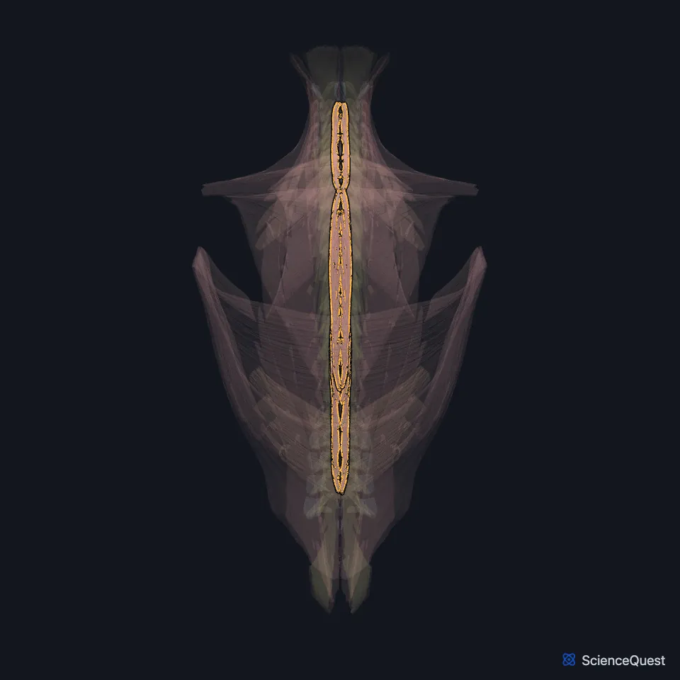
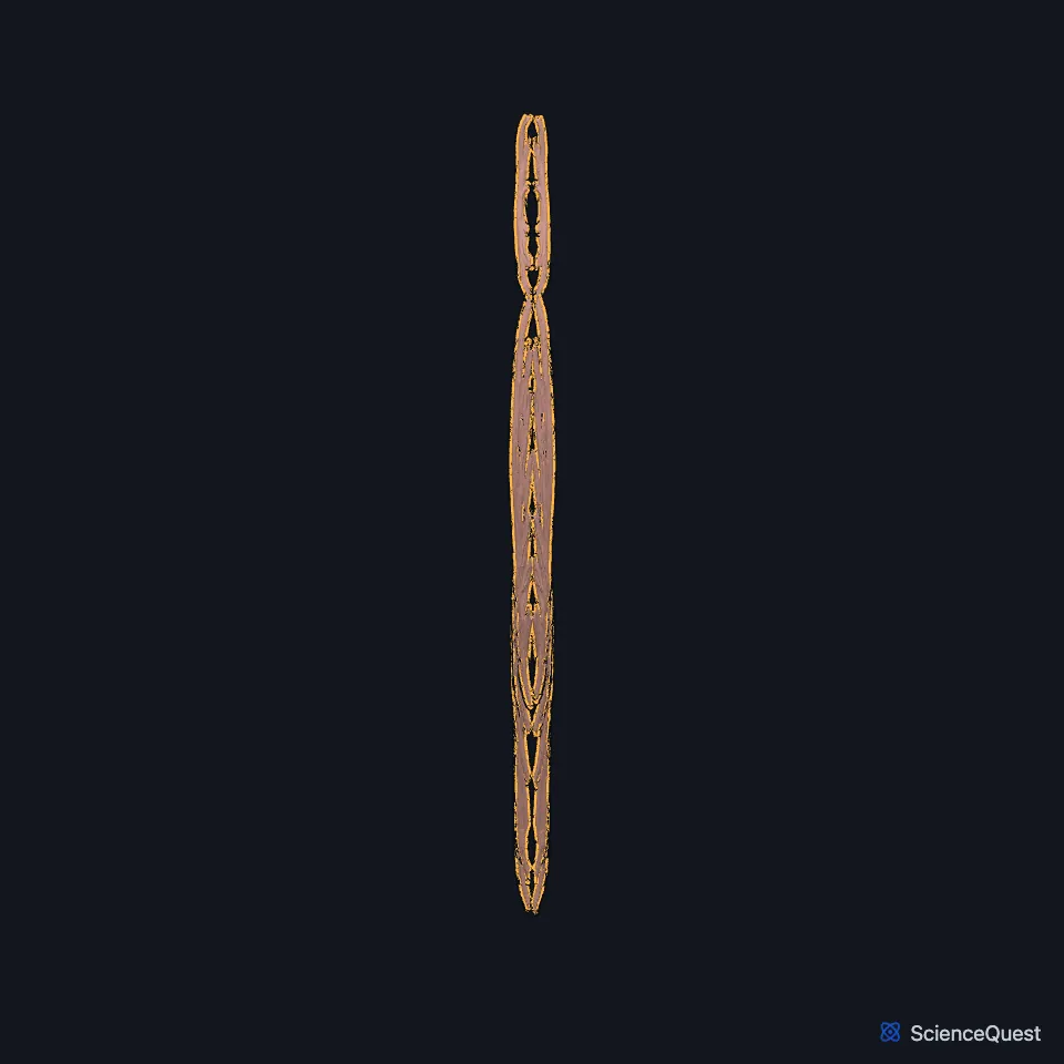
Where is the spinalis?
Gray’s 1918 text describes the erector spinae splitting in the upper lumbar region into three columns: the iliocostalis outermost, the longissimus in the middle and the spinalis innermost. (Gray 1918, 6a. Deep Muscles of the Back)
In the thoracic region the spinalis thoracis is the most medial part of the erector spinae, and it blends with the longissimus thoracis on its outer side. (StatPearls, Back, Muscles)
What are the parts of the spinalis?
It has three parts: the spinalis thoracis, the spinalis cervicis and the spinalis capitis. Gray’s 1918 text calls the thoracic part the spinalis dorsi. (StatPearls, Back, Muscles; Gray 1918, 6a. Deep Muscles of the Back)
The spinalis thoracis runs from the spines of T11 to L2 to the spines of the upper thoracic vertebrae. Gray’s 1918 text puts its insertions on four to eight upper thoracic spines and says it is difficult to separate from the longissimus as a muscle of its own. (StatPearls, Back, Muscles; Gray 1918, 6a. Deep Muscles of the Back)
The spinalis cervicis runs from the ligamentum nuchae and the spine of C7 to the spines of the axis and C3 to C4. StatPearls describes the cervicis and capitis as ill-defined and underdeveloped, and absent in some people. (StatPearls, Back, Muscles)
StatPearls describes the spinalis capitis as usually a few fibers of the semispinalis capitis. Gray’s 1918 text calls it usually inseparable from that muscle, and in its account of the semispinalis capitis gives the name to the muscle’s inner part, usually more or less distinct from the rest. (Gray 1918, 6a. Deep Muscles of the Back; StatPearls, Back, Muscles)
What does the spinalis do?
As part of the erector spinae, it extends the back when both sides contract, and bends and rotates the spine when one side contracts. The group also holds posture and controls bending forward. (StatPearls, Back and Back, Muscles)
What covers, supplies and underlies the spinalis?
The thoracolumbar fascia, the serratus posterior inferior, the rhomboids and the splenius muscles cover the erector spinae. (StatPearls, Back, Muscles)
Beneath it lies the semispinalis thoracis, which Gray’s 1918 text calls the semispinalis dorsi and describes as joined to it. (Gray 1918, 6a. Deep Muscles of the Back)
Like the other intrinsic back muscles, it takes its nerves from the dorsal rami of the spinal nerves. (StatPearls, Back, Muscles)
| Feature | Detail and source |
|---|---|
| Parts | Spinalis thoracis, cervicis and capitis (StatPearls, Back, Muscles; Gray 1918, 6a. Deep Muscles of the Back) |
| Origin | Spines of T11 to L2 (thoracis); ligamentum nuchae and the C7 spine (cervicis) (StatPearls, Back, Muscles) |
| Insertion | Spines of the upper thoracic vertebrae (thoracis); spines of the axis and C3 to C4 (cervicis) (StatPearls, Back, Muscles) |
| Action | As part of the erector spinae: extends the spine when both sides act; bends it sideways and rotates it when one side acts (StatPearls, Back) |
| Nerve | Dorsal rami of the spinal nerves (StatPearls, Back, Muscles) |
Touches in this model
Measured on the BodyParts3D meshes: surfaces within 2 mm of the spinalis, nearest first, 12 of 52.
- Left longissimus thoracis in contact
- Left multifidus in contact
- Left semispinalis cervicis in contact
- Left semispinalis thoracis in contact
- Left serratus posterior inferior in contact
- Left splenius capitis in contact
- Left splenius cervicis in contact
- Right longissimus thoracis in contact
- Right multifidus in contact
- Right rhomboid major in contact
- Right semispinalis cervicis in contact
- Right semispinalis thoracis in contact
Sources
Sources read
- StatPearls, Anatomy, Back. Robert J. Modes; Sevda Lafci Fahrioglu. Last update . CC BY-NC-ND 4.0. Read .
- StatPearls, Anatomy, Back, Muscles. Brandi Henson; Bhavana Kadiyala; Mary Ann Edens. Last update . CC BY-NC-ND 4.0. Read .
- Gray’s Anatomy of the Human Body, 20th US edition (1918), IV. Myology, 6a. The Deep Muscles of the Back (1918 terms: Sacrospinalis for erector spinae, Iliocostalis dorsi for iliocostalis thoracis, Longissimus dorsi for longissimus thoracis). Henry Gray. Public domain. Read .
The values are facts and free to use; this page’s selection and presentation are © 2026 ScienceQuest.