Flexor digitorum longus
The flexor digitorum longus is a muscle of the deep posterior compartment, on the tibial side of the leg. It flexes the four lateral toes and helps plantarflex the ankle. (Gray’s Anatomy (1918), Posterior Crural Muscles, deep group; StatPearls, Leg Posterior Compartment)
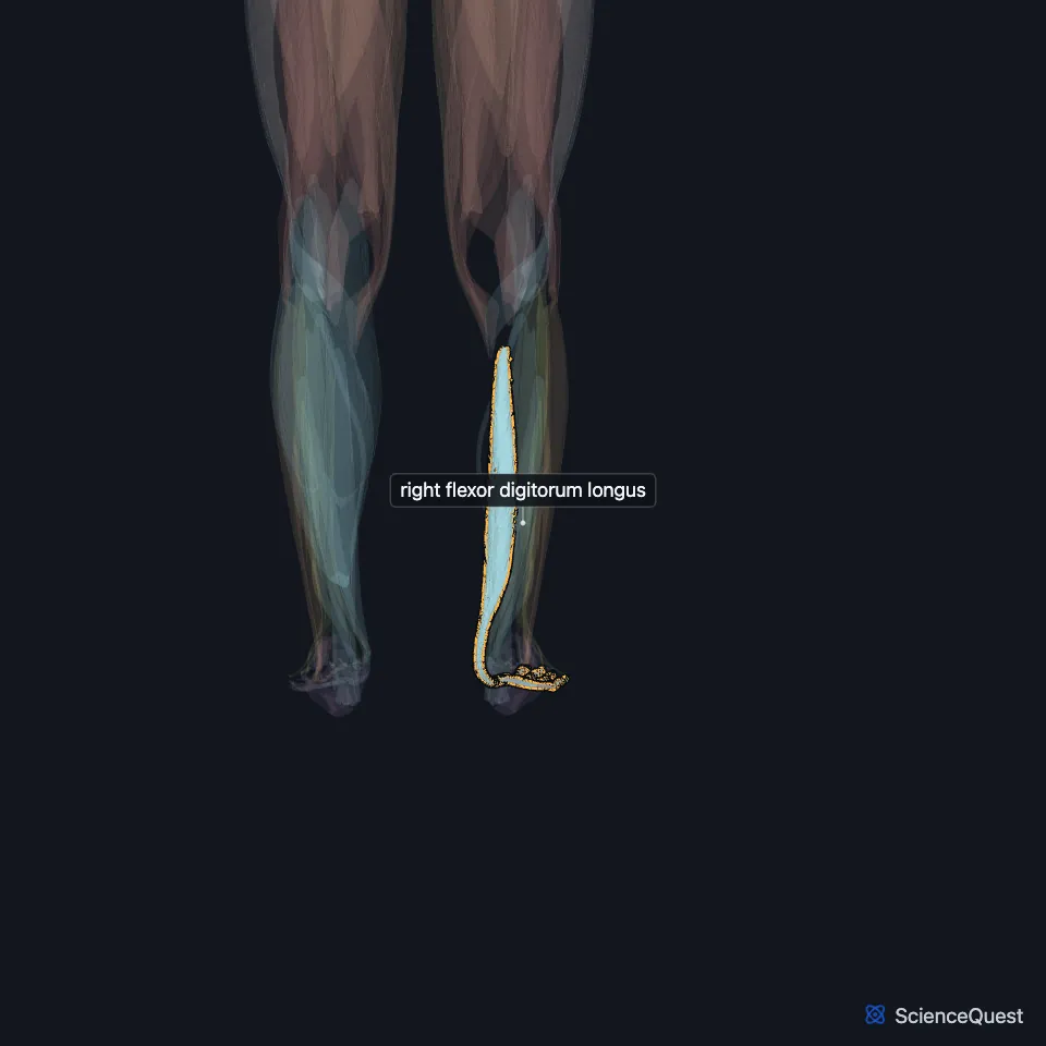
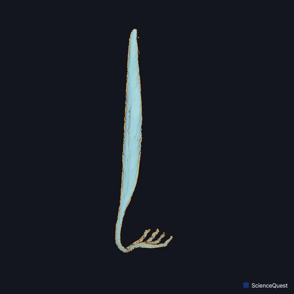
Open it in the Lower Limb Muscles Explorer See it alone in 3D
Where is the flexor digitorum longus?
It lies on the medial side of the back of the leg. Thin and pointed at its origin, it grows larger as it descends. (StatPearls, Calf; Gray’s Anatomy (1918), Posterior Crural Muscles, deep group)
It arises from the back of the tibia below the soleal line, medial to the tibial origin of the tibialis posterior, and from the fascia over that muscle. (StatPearls, Tibial Nerve; Gray’s Anatomy (1918), Posterior Crural Muscles, deep group)
What path does its tendon take?
Its tendon passes behind the medial malleolus in a groove it shares with the tibialis posterior tendon, each tendon in its own compartment and sheath. In the lower fourth of the leg the tibialis posterior tendon passes in front of it. (Gray’s Anatomy (1918), Posterior Crural Muscles, deep group)
In the sole it crosses below the flexor hallucis longus tendon and takes a strong slip from it. It then divides into four tendons that insert on the bases of the distal phalanges of the second to fifth toes. (Gray’s Anatomy (1918), Posterior Crural Muscles, deep group; StatPearls, Tibial Nerve)
Each of the four tendons passes through an opening in the matching tendon of the flexor digitorum brevis. The lumbricals of the foot arise from these tendons and have no bony origin. (Gray’s Anatomy (1918), Posterior Crural Muscles, deep group; StatPearls, Arches of the Foot)
What does the flexor digitorum longus do?
It flexes the second to fifth toes at the distal and proximal interphalangeal and metatarsophalangeal joints, and plantarflexes the ankle. (StatPearls, Tibial Nerve)
It helps maintain both the medial and lateral longitudinal arches, acting as a tie beam. (StatPearls, Leg Posterior Compartment and Arches of the Foot)
Its tendons run at a slant, so on their own they would draw the toes medially. Gray’s 1918 credits the quadratus plantae, inserted on the lateral side of the tendon, with pulling it back to the middle line of the foot. (Gray’s Anatomy (1918), Posterior Crural Muscles, deep group)
What supplies the flexor digitorum longus?
The tibial nerve supplies it. The nerve passes behind the medial malleolus between its tendon and the flexor hallucis longus tendon, in the tarsal tunnel. (StatPearls, Tibial Nerve)
Does the flexor digitorum longus vary?
An accessory flexor digitorum longus occurs in about 15% of people. Gray’s 1918 describes it arising from the fibula, tibia or deep fascia and joining the long flexor tendon after it passes under the laciniate ligament, another name for the flexor retinaculum. (StatPearls, Leg Posterior Compartment and Tibialis Posterior Muscle; Gray’s Anatomy (1918), Posterior Crural Muscles, deep group)
Where the sources differ
Sources disagree on the extent of its tibial origin: the middle third of the back of the tibia in the StatPearls posterior compartment article, but from just below what Gray’s 1918 calls the popliteal line to within 7 or 8 cm of the lower end in Gray’s. (StatPearls, Leg Posterior Compartment; Gray’s Anatomy (1918), Posterior Crural Muscles, deep group)
Sources give different roots for its nerve: S1 and S2 in the StatPearls popliteus article, the fifth lumbar and first sacral nerves in Gray’s 1918. (StatPearls, Popliteus Muscle; Gray’s Anatomy (1918), Posterior Crural Muscles, deep group)
| Feature | Detail and source |
|---|---|
| Origin | Back of the tibia, below the soleal line (StatPearls, Tibial Nerve) |
| Insertion | Bases of the distal phalanges of toes 2 to 5 (StatPearls, Tibial Nerve) |
| Action | Flexes toes 2 to 5; plantarflexes the ankle (StatPearls, Tibial Nerve) |
| Nerve | Tibial nerve (StatPearls, Tibial Nerve) |
Touches in this model
Measured on the BodyParts3D meshes: surfaces within 2 mm of the flexor digitorum longus, nearest first, 12 of 33.
- First lumbrical of right foot in contact
- Flexor digiti minimi brevis of right foot in contact
- Fourth lumbrical of right foot in contact
- Oblique head of right adductor hallucis in contact
- Right abductor hallucis in contact
- Right flexor accessorius in contact
- Right flexor digitorum brevis in contact
- Right flexor hallucis longus in contact
- Right soleus in contact
- Right tibialis posterior in contact
- Second lumbrical of right foot in contact
- Third lumbrical of right foot in contact
Sources
Sources read
- Anatomy of the Human Body (Gray’s Anatomy), 20th US edition, 1918, Myology, The Muscles and Fasciae of the Leg, 2. The Posterior Crural Muscles, the deep group, pages 483 to 486; Internet Archive scan of the 1918 Lea and Febiger printing, terminology of 1918. Henry Gray; edited by Warren H. Lewis. Public domain. Read .
- StatPearls, Anatomy, Bony Pelvis and Lower Limb: Leg Posterior Compartment. Rami Abuqubo; Steven B. Graefe; James La Rose. Last update . CC BY-NC-ND 4.0. Read .
- StatPearls, Anatomy, Bony Pelvis and Lower Limb: Calf. Justin T. Binstead; Matthew A. Varacallo. Last update . CC BY-NC-ND 4.0. Read .
- StatPearls, Anatomy, Bony Pelvis and Lower Limb: Tibial Nerve. Sohil S. Desai; James La Rose. Last update . CC BY-NC-ND 4.0. Read .
- StatPearls, Anatomy, Bony Pelvis and Lower Limb: Arches of the Foot. Hiteshkumar M. Chauhan; Muhammad Taqi. Last update . CC BY-NC-ND 4.0. Read .
- StatPearls, Anatomy, Bony Pelvis and Lower Limb: Tibialis Posterior Muscle. Nicholas M. Corcoran; Matthew A. Varacallo. Last update . CC BY-NC-ND 4.0. Read .
- StatPearls, Anatomy, Bony Pelvis and Lower Limb: Popliteus Muscle. Scott Hyland; Matthew A. Varacallo. Last update . CC BY-NC-ND 4.0. Read .
The values are facts and free to use; this page’s selection and presentation are © 2026 ScienceQuest.