Flexor digitorum brevis
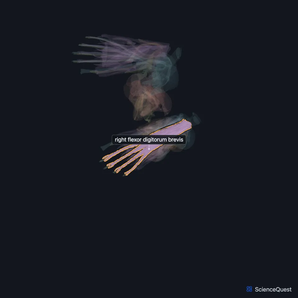
The flexor digitorum brevis lies in the middle of the sole, in its first muscle layer, and flexes the four lesser toes. (Gray’s Anatomy (1918), Plantar Muscles of the Foot; StatPearls, Foot Muscles)
Open it in the Lower Limb Muscles Explorer See it alone in 3D
Where is the flexor digitorum brevis?
It sits in the superficial central compartment of the sole with the four lumbricals and the tendons of the flexor digitorum longus. A thin layer of fascia separates its deep surface from the lateral plantar vessels and nerves. (StatPearls, Foot Muscles; Gray’s Anatomy (1918), Plantar Muscles of the Foot)
The abductor hallucis runs along the medial border of the sole and the abductor digiti minimi along the lateral border, and septa between them and this muscle give origin to all three. (Gray’s Anatomy (1918), Plantar Muscles of the Foot)
What does the flexor digitorum brevis attach to?
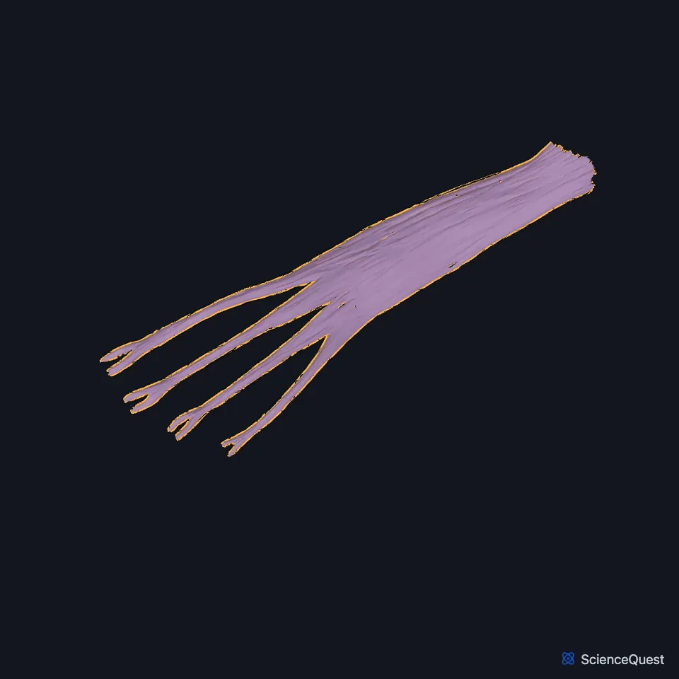
It arises from the medial process of the tuberosity of the calcaneus, the central part of the plantar aponeurosis and the septa beside it. It then divides into four tendons, one for each lesser toe. (Gray’s Anatomy (1918), Plantar Muscles of the Foot)
At the base of each proximal phalanx a tendon splits to let the long flexor tendon through. The two slips then rejoin to form a grooved channel for that tendon, divide a second time and insert on the sides of the middle phalanx. (Gray’s Anatomy (1918), Plantar Muscles of the Foot)
What does the flexor digitorum brevis do?
It flexes the middle phalanges of the lesser toes on the proximal ones and, as it keeps acting, flexes the proximal phalanges too. (Gray’s Anatomy (1918), Plantar Muscles of the Foot; StatPearls, Foot Muscles)
Which nerve supplies the flexor digitorum brevis, and what lies under it?
The medial plantar nerve supplies it, and the medial plantar artery brings its blood. (StatPearls, Foot Muscles)
Under the first layer, in the second, the flexor accessorius joins the tendon of the flexor digitorum longus. The lateral plantar vessels and nerve separate it from the first-layer muscles. (Gray’s Anatomy (1918), Plantar Muscles of the Foot)
| Feature | Detail and source |
|---|---|
| Origin | Calcaneal tuberosity; plantar aponeurosis; intermuscular septa (StatPearls, Foot Muscles) |
| Insertion | Middle phalanges of the second to fifth toes (StatPearls, Foot Muscles) |
| Action | Flexes the second to fifth toes (StatPearls, Foot Muscles) |
| Nerve | Medial plantar nerve (StatPearls, Foot Muscles) |
Touches in this model
Measured on the BodyParts3D meshes: surfaces within 2 mm of the flexor digitorum brevis, nearest first, 12 of 17.
- Abductor digiti minimi of right foot in contact
- Fourth lumbrical of right foot in contact
- Right abductor hallucis in contact
- Right flexor accessorius in contact
- Right flexor digitorum longus in contact
- Middle phalanx of right second toe in contact
- Middle phalanx of right third toe in contact
- Proximal phalanx of right little toe in contact
- Right calcaneus in contact
- Skin in contact
- Middle phalanx of right little toe 0.1 mm away
- Right long plantar ligament 0.1 mm away
Sources
Sources read
- Anatomy of the Human Body (Gray’s Anatomy), 20th US edition, 1918, Myology, The Muscles and Fasciae of the Foot, 2. The Plantar Muscles of the Foot, pages 490 to 496; Internet Archive scan of the 1918 Lea and Febiger printing, terminology of 1918. Henry Gray; edited by Warren H. Lewis. Public domain. Read .
- StatPearls, Anatomy, Bony Pelvis and Lower Limb, Foot Muscles. Ryan K. Card; Bruno Bordoni. Last update . CC BY-NC-ND 4.0. Read .
The values are facts and free to use; this page’s selection and presentation are © 2026 ScienceQuest.