Extensor digitorum brevis
The extensor digitorum brevis is a broad, thin muscle on the dorsum of the foot that helps extend the toes. (Gray’s Anatomy (1918), Dorsal Muscle of the Foot; StatPearls, Foot Muscles)
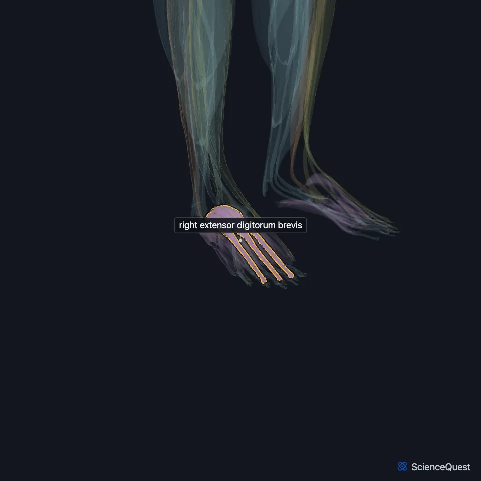
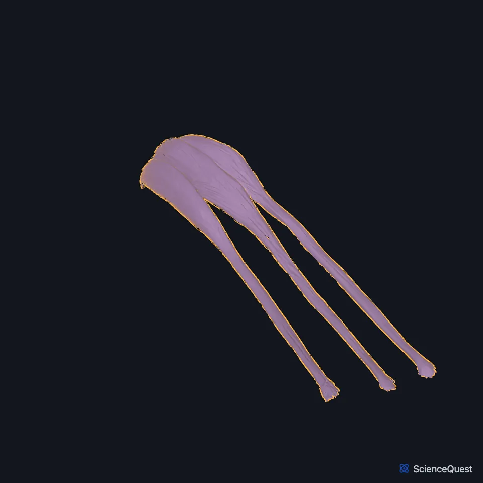
Open it in the Lower Limb Muscles Explorer See it alone in 3D
Where is the extensor digitorum brevis?
It arises from the front of the upper and lateral surfaces of the calcaneus and runs obliquely across the dorsum of the foot toward the toes. (Gray’s Anatomy (1918), Dorsal Muscle of the Foot)
What does the extensor digitorum brevis attach to?
StatPearls gives its origin as the dorsolateral surface of the calcaneus, the inferior extensor retinaculum and the talocalcaneal interosseous ligament. Its tendons join the lateral sides of the extensor digitorum longus tendons to the second, third and fourth toes. (StatPearls, Foot Muscles; Gray’s Anatomy (1918), Dorsal Muscle of the Foot)
Its tendons vary. Gray’s 1918 records as few as two, a doubled tendon, or an extra slip to the little toe. (Gray’s Anatomy (1918), Dorsal Muscle of the Foot)
What does the extensor digitorum brevis do?
It extends the second to fourth toes. Gray’s 1918 explains that its oblique line of pull offsets the oblique pull of the long extensor, so the toes extend evenly when both act. (StatPearls, Foot Muscles; Gray’s Anatomy (1918), Dorsal Muscle of the Foot)
Which nerve and arteries supply the extensor digitorum brevis?
The deep fibular nerve supplies it. StatPearls also calls it the deep peroneal nerve, the name Gray’s 1918 uses. The fibular, anterior tibial and dorsalis pedis arteries bring its blood. (StatPearls, Foot Muscles and Calf Deep Peroneal Nerve; Gray’s Anatomy (1918), Dorsal Muscle of the Foot)
In up to 28% of people an accessory deep fibular nerve, usually from the superficial fibular nerve, supplies part or all of the muscle on one or both sides. (StatPearls, Calf Deep Peroneal Nerve)
The lateral terminal branch of the deep fibular nerve runs beneath it and supplies both it and the extensor hallucis brevis. (StatPearls, Calf Deep Peroneal Nerve)
Where the sources differ
Sources disagree on what belongs to the muscle. Gray’s 1918 gives it four tendons, the most medial and largest going to the great toe and often described as a separate muscle; StatPearls treats the extensor hallucis brevis as its own muscle and gives the extensor digitorum brevis the tendons to toes 2 to 4. (Gray’s Anatomy (1918), Dorsal Muscle of the Foot; StatPearls, Foot Muscles)
Sources name different ligaments in its origin. Besides the dorsolateral calcaneus, StatPearls gives the inferior extensor retinaculum and the talocalcaneal interosseous ligament. Gray’s 1918 gives the lateral talocalcanean ligament and the common limb of the cruciate crural ligament, a 1918 term. (StatPearls, Foot Muscles; Gray’s Anatomy (1918), Dorsal Muscle of the Foot)
| Feature | Detail and source |
|---|---|
| Origin | Dorsolateral calcaneus; inferior extensor retinaculum; talocalcaneal interosseous ligament (sources differ; see below) (StatPearls, Foot Muscles) |
| Insertion | Extensor digitorum longus tendons to the second, third and fourth toes (StatPearls, Foot Muscles) |
| Action | Extends the second to fourth toes (StatPearls, Foot Muscles) |
| Nerve | Deep fibular nerve (StatPearls, Foot Muscles) |
Touches in this model
Measured on the BodyParts3D meshes: surfaces within 2 mm of the extensor digitorum brevis, nearest first, 12 of 19.
- Right extensor digitorum longus in contact
- Right extensor hallucis brevis in contact
- Right fibularis tertius in contact
- Set of dorsal interossei of right foot in contact
- Middle phalanx of right fourth toe in contact
- Middle phalanx of right second toe in contact
- Middle phalanx of right third toe in contact
- Proximal phalanx of right fourth toe in contact
- Proximal phalanx of right second toe in contact
- Right calcaneus in contact
- Right cuboid bone in contact
- Right fourth metatarsal bone in contact
Sources
Sources read
- Anatomy of the Human Body (Gray’s Anatomy), 20th US edition, 1918, Myology, The Muscles and Fasciae of the Foot, 1. The Dorsal Muscle of the Foot, page 490; Internet Archive scan of the 1918 Lea and Febiger printing, terminology of 1918. Henry Gray; edited by Warren H. Lewis. Public domain. Read .
- StatPearls, Anatomy, Bony Pelvis and Lower Limb, Foot Muscles. Ryan K. Card; Bruno Bordoni. Last update . CC BY-NC-ND 4.0. Read .
- StatPearls, Anatomy, Bony Pelvis and Lower Limb: Calf Deep Peroneal Nerve (Deep Fibular Nerve). Lane L. Palmisano; Marjorie V. Launico. Last update . CC BY-NC-ND 4.0. Read .
The values are facts and free to use; this page’s selection and presentation are © 2026 ScienceQuest.