Extensor digitorum longus
The extensor digitorum longus is one of the four muscles of the anterior compartment of the leg. It extends the second to fifth toes and helps dorsiflex the ankle. (StatPearls, Leg Anterior Compartment)
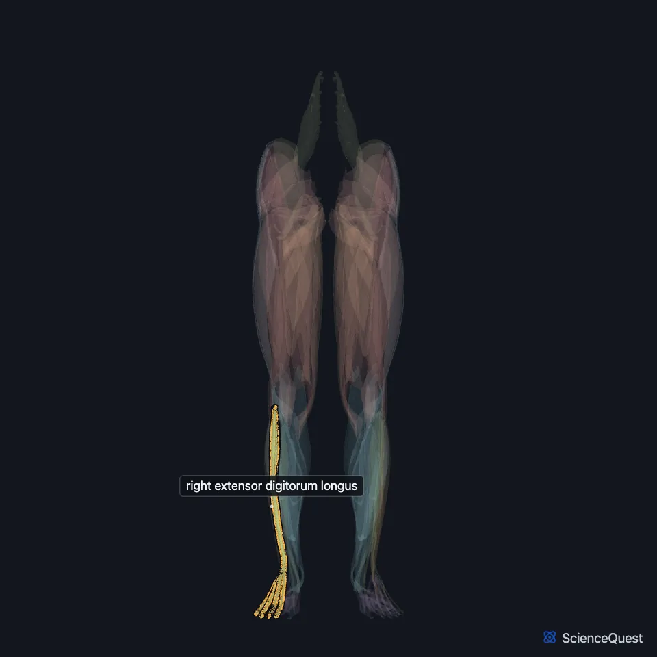
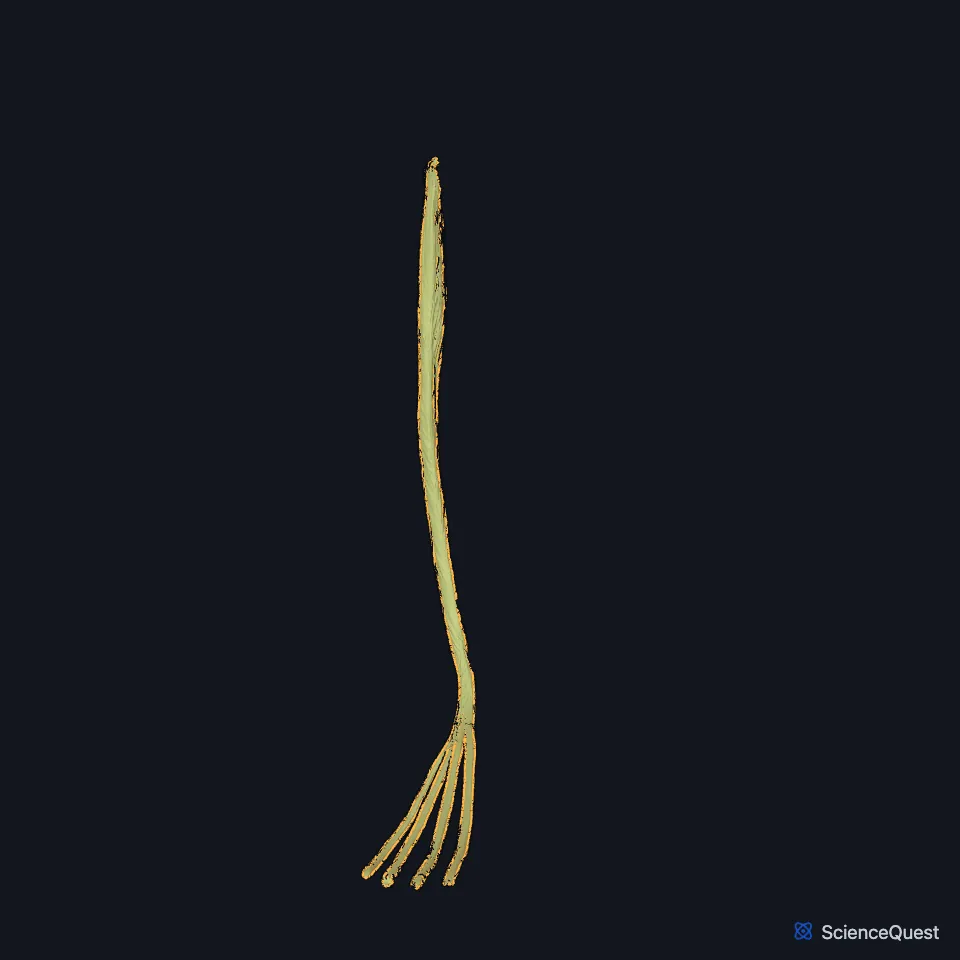
Open it in the Lower Limb Muscles Explorer See it alone in 3D
Where is the extensor digitorum longus?
It runs down the lateral part of the front of the leg and forms the central, superficial part of the anterior compartment. An intermuscular septum separates it from the tibialis anterior on its medial side. (StatPearls, Leg Anterior Compartment; Gray’s Anatomy (1918), Anterior Crural Muscles)
It arises from the lateral condyle of the tibia, the anterior surface of the fibula and the interosseous membrane. Gray’s 1918 adds fibers from the deep surface of the fascia of the leg. (StatPearls, Leg Anterior Compartment; Gray’s Anatomy (1918), Anterior Crural Muscles)
How is the extensor digitorum longus built?
Its tendon passes under the transverse and cruciate crural ligaments, as Gray’s 1918 names them, and divides into four slips that run forward on the top of the foot. The slips end in the dorsal expansions of the second to fifth toes, reaching their middle and distal phalanges. (Gray’s Anatomy (1918), Anterior Crural Muscles; StatPearls, Leg Anterior Compartment)
Opposite the metatarsophalangeal joints, a tendon of the extensor digitorum brevis joins the lateral side of the slips to the second, third and fourth toes. Each slip also takes a fibrous expansion from the interossei and a lumbrical, then spreads into an aponeurosis over the first, or proximal, phalanx. (Gray’s Anatomy (1918), Anterior Crural Muscles)
At the next joint the aponeurosis divides into three slips. The middle slip inserts on the base of the second phalanx, and the two side slips reunite and insert on the base of the third, the middle and distal phalanges in current terms. (Gray’s Anatomy (1918), Anterior Crural Muscles; StatPearls, Leg Anterior Compartment)
The fibularis tertius often arises as its lower extension, and Gray’s 1918 calls that muscle its fifth tendon. (StatPearls, Leg Anterior Compartment; Gray’s Anatomy (1918), Anterior Crural Muscles)
What does the extensor digitorum longus do?
With the other anterior compartment muscles it lifts the foot clear of the ground in the swing phase of walking. The same group steadies the foot at heel strike and through stance. (StatPearls, Leg Anterior Compartment)
In standing, these front leg muscles pull from their attachments below to hold the leg bones upright and strengthen the ankle joint. (Gray’s Anatomy (1918), Anterior Crural Muscles)
What supplies the extensor digitorum longus?
The deep fibular nerve supplies it and the other three muscles of the compartment. Gray’s 1918 traces the supply of these muscles to the fourth and fifth lumbar and first sacral nerves. (StatPearls, Calf Deep Peroneal Nerve; Gray’s Anatomy (1918), Anterior Crural Muscles)
The anterior tibial artery, a branch of the popliteal artery, supplies the compartment. In the upper leg the artery and the deep fibular nerve run between this muscle and the tibialis anterior. (StatPearls, Leg Anterior Compartment; Gray’s Anatomy (1918), Anterior Crural Muscles)
Does the extensor digitorum longus vary?
Accessory slips and extra links to nearby tendons occur. Gray’s 1918 records doubled tendons to the second and fifth toes and extra slips to the metatarsals, the short extensor or an interosseous muscle. (StatPearls, Leg Anterior Compartment; Gray’s Anatomy (1918), Anterior Crural Muscles)
| Feature | Detail and source |
|---|---|
| Origin | Lateral condyle of the tibia; anterior surface of the fibula; interosseous membrane (StatPearls, Leg Anterior Compartment) |
| Insertion | Dorsal expansions of toes 2 to 5, to the middle and distal phalanges (StatPearls, Leg Anterior Compartment) |
| Action | Extends toes 2 to 5; assists dorsiflexion of the ankle (StatPearls, Leg Anterior Compartment) |
| Nerve | Deep fibular (deep peroneal) nerve (StatPearls, Leg Anterior Compartment) |
Touches in this model
Measured on the BodyParts3D meshes: surfaces within 2 mm of the extensor digitorum longus, nearest first, 12 of 32.
- Right extensor digitorum brevis in contact
- Right extensor hallucis longus in contact
- Right fibularis brevis in contact
- Right fibularis longus in contact
- Right fibularis tertius in contact
- Right iliotibial tract in contact
- Right tibialis anterior in contact
- Right tibialis posterior in contact
- Distal phalanx of right little toe in contact
- Interosseous membrane of right leg in contact
- Middle phalanx of right fourth toe in contact
- Middle phalanx of right second toe in contact
Sources
Sources read
- StatPearls, Anatomy, Bony Pelvis and Lower Limb: Leg Anterior Compartment. Bradley Lezak; Marjorie V. Launico. Last update . CC BY-NC-ND 4.0. Read .
- Anatomy of the Human Body (Gray’s Anatomy), 20th US edition, 1918, Myology, The Muscles and Fasciae of the Leg, 1. The Anterior Crural Muscles, pages 480 to 482; Internet Archive scan of the 1918 Lea and Febiger printing, terminology of 1918. Henry Gray; edited by Warren H. Lewis. Public domain. Read .
- StatPearls, Anatomy, Bony Pelvis and Lower Limb: Calf Deep Peroneal Nerve (Deep Fibular Nerve). Lane L. Palmisano; Marjorie V. Launico. Last update . CC BY-NC-ND 4.0. Read .
The values are facts and free to use; this page’s selection and presentation are © 2026 ScienceQuest.