Fibularis brevis
The fibularis brevis, also called the peroneus brevis, is the shorter of the two muscles of the lateral compartment of the leg and lies under the fibularis longus. It everts the foot and, to a lesser extent, plantarflexes the ankle. (StatPearls, Foot Peroneus Brevis Muscle)
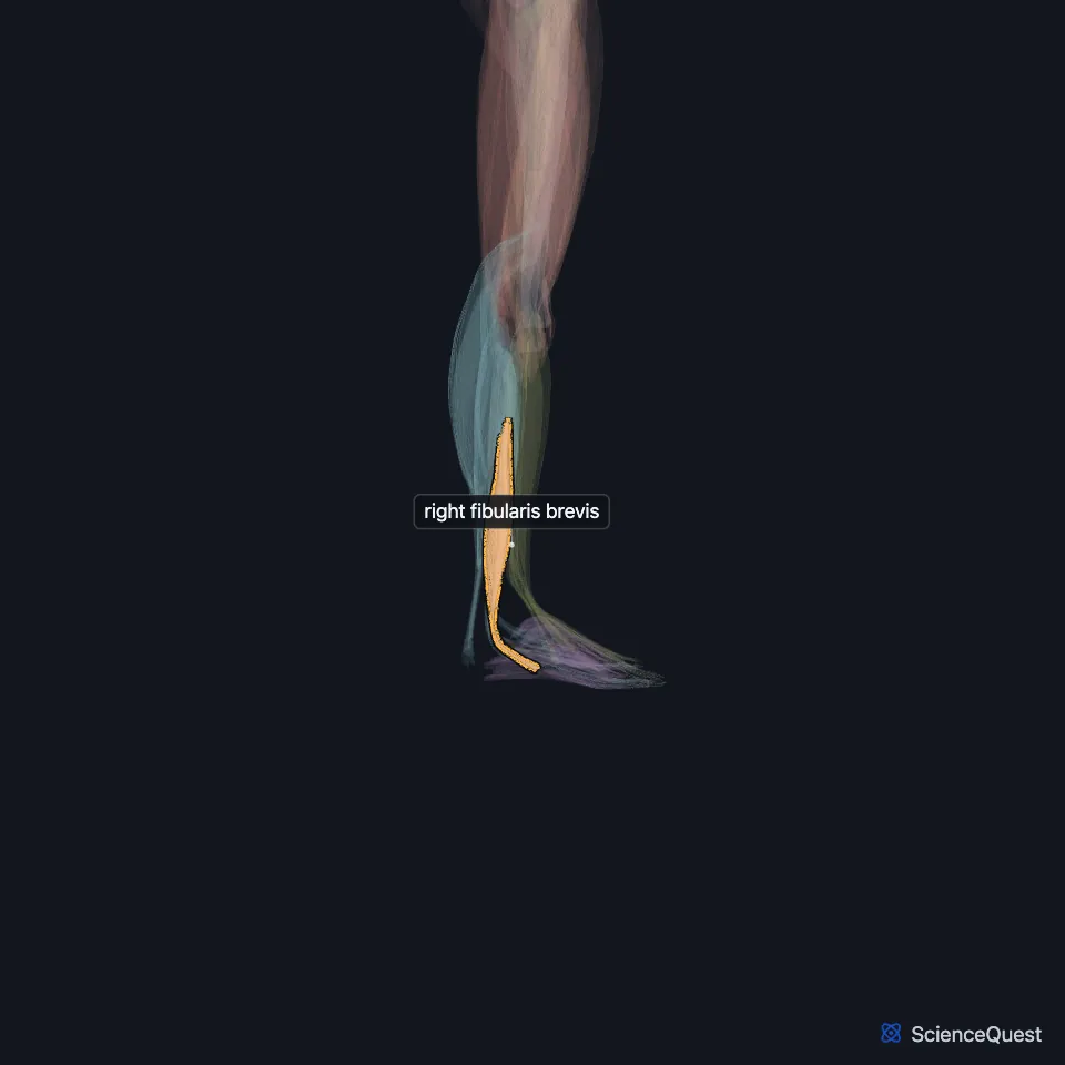
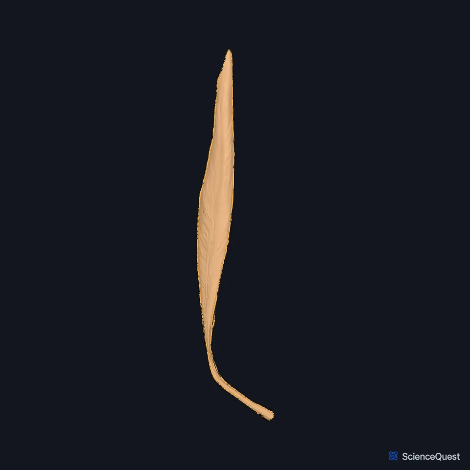
Open it in the Lower Limb Muscles Explorer See it alone in 3D
Where is the fibularis brevis?
It lies deep and medial to the fibularis longus and arises from the lower two-thirds of the lateral surface of the fibula and the anterior intermuscular septum. (StatPearls, Leg Lateral Compartment)
It is shorter than the fibularis longus but has the thicker belly. (StatPearls, Foot Peroneus Brevis Muscle)
What path does its tendon take?
Its tendon begins about 2 to 4 cm above the tip of the lateral malleolus. Behind the malleolus it runs in front of the longus tendon, the two sharing one synovial sheath under the superior fibular retinaculum. (StatPearls, Foot Peroneus Brevis Muscle and Leg Lateral Compartment)
On the side of the calcaneus it passes above the fibular trochlea (peroneal tubercle), with the longus tendon below. It inserts on the lateral side of the tuberosity at the base of the fifth metatarsal. (StatPearls, Foot Peroneus Brevis Muscle and Leg Lateral Compartment; Gray’s Anatomy (1918), Calcaneus, p. 263)
In cadaver studies the tendon averages 79.6 mm long, 4.46 mm wide at its midpoint and 14.85 mm wide at its insertion. (StatPearls, Foot Peroneus Brevis Muscle)
What does the fibularis brevis do?
Its insertion on the lateral side of the foot lets it evert the foot, tilting the sole away from the midline. Its insertion close to the ankle lets it plantarflex too, with the ankle joint as the fulcrum for both movements. (StatPearls, Foot Peroneus Brevis Muscle)
With the fibularis tertius and the tibialis anterior it gives suspensory support to the transverse arch of the foot. (StatPearls, Arches of the Foot)
What supplies and holds the fibularis brevis?
A muscular branch of the superficial fibular nerve supplies it. Its parent, the common fibular nerve, winds around the neck of the fibula, and Gray’s 1918 calls it the only nerve of the lower limb you can find by touch there. (StatPearls, Foot Peroneus Brevis Muscle and Calf Peroneus Longus Muscle; Gray’s Anatomy (1918), Surface Anatomy of the Lower Extremity)
The superior and inferior fibular retinacula hold its tendon and the longus tendon at the ankle and stop them subluxing. (StatPearls, Foot Peroneus Brevis Muscle)
Even so, the two fibular tendons are the ankle tendons that dislocate most often, because the groove behind the fibula is shallow in some people. (StatPearls, Foot Peroneus Brevis Muscle)
Does the fibularis brevis vary?
The peroneus quartus, the commonest variant muscle of the lateral compartment, occurs in 5% to 20% of people. In most of them it arises from the fibularis brevis and inserts on the calcaneus. (StatPearls, Foot Peroneus Brevis Muscle)
Its insertion varies as well: in one classification, 70% of people have a single insertion on the fifth metatarsal tuberosity, and the rest add an accessory band. (StatPearls, Leg Lateral Compartment)
Where the sources differ
Sources give different spinal roots for its nerve. The StatPearls fibularis brevis article gives L5 to S2, the StatPearls fibularis longus article gives both L5 to S2 and L4 to S1 for the superficial fibular nerve, and Gray’s 1918 gives the fourth and fifth lumbar and first sacral nerves. (StatPearls, Foot Peroneus Brevis Muscle and Calf Peroneus Longus Muscle; Gray’s Anatomy (1918), Lateral Crural Muscles)
Sources disagree on its blood supply: the StatPearls fibularis brevis article names muscular branches of the fibular artery, while the lateral compartment article has the anterior tibial artery supply the upper fibular muscles and the fibular artery their lower parts. (StatPearls, Foot Peroneus Brevis Muscle and Leg Lateral Compartment)
| Feature | Detail and source |
|---|---|
| Origin | Lower two-thirds of the lateral fibula; anterior intermuscular septum (StatPearls, Leg Lateral Compartment) |
| Insertion | Lateral side of the tuberosity at the base of the fifth metatarsal (StatPearls, Leg Lateral Compartment) |
| Action | Everts the foot; plantarflexes the ankle to a lesser extent (StatPearls, Foot Peroneus Brevis Muscle) |
| Nerve | Superficial fibular (superficial peroneal) nerve (StatPearls, Foot Peroneus Brevis Muscle) |
Touches in this model
Measured on the BodyParts3D meshes: surfaces within 2 mm of the fibularis brevis, nearest first, 12 of 13.
- Abductor digiti minimi of right foot in contact
- Right extensor digitorum longus in contact
- Right fibularis longus in contact
- Right flexor hallucis longus in contact
- Right soleus in contact
- Right calcaneus in contact
- Right fibula in contact
- Right fifth metatarsal bone in contact
- Right talus in contact
- Right tibia in contact
- Skin in contact
- Right cuboid bone 1.1 mm away
Sources
Sources read between and
- StatPearls, Anatomy, Bony Pelvis and Lower Limb, Foot Peroneus Brevis Muscle. Hajira Basit; Jamie Shah; Marco A. Siccardi. Last update . CC BY-NC-ND 4.0. Read .
- StatPearls, Anatomy, Bony Pelvis and Lower Limb: Leg Lateral Compartment. Irfan A. Khan; Navid Mahabadi; Anthony D’Abarno; Matthew A. Varacallo. Last update . CC BY-NC-ND 4.0. Read .
- Gray’s Anatomy of the Human Body, 20th US edition (1918), edited by Warren H. Lewis, Osteology, The Calcaneus (pp. 263 to 266). Henry Gray; Warren H. Lewis (editor). Public domain. Read .
- StatPearls, Anatomy, Bony Pelvis and Lower Limb: Arches of the Foot. Hiteshkumar M. Chauhan; Muhammad Taqi. Last update . CC BY-NC-ND 4.0. Read .
- StatPearls, Anatomy, Bony Pelvis and Lower Limb: Calf Peroneus Longus Muscle. Bradley Lezak; Matthew A. Varacallo. Last update . CC BY-NC-ND 4.0. Read .
- Anatomy of the Human Body (Gray’s Anatomy), 20th US edition, 1918, Surface Anatomy and Surface Markings, Surface Anatomy of the Lower Extremity, Nerves, page 1342; Internet Archive scan of the 1918 Lea and Febiger printing, terminology of 1918. Henry Gray; edited by Warren H. Lewis. Public domain. Read .
- Anatomy of the Human Body (Gray’s Anatomy), 20th US edition, 1918, Myology, The Muscles and Fasciae of the Leg, 3. The Lateral Crural Muscles, pages 486 to 488; Internet Archive scan of the 1918 Lea and Febiger printing, terminology of 1918. Henry Gray; edited by Warren H. Lewis. Public domain. Read .
The values are facts and free to use; this page’s selection and presentation are © 2026 ScienceQuest.