Fibularis longus
The fibularis longus, also called the peroneus longus, is the longer and more superficial of the two muscles of the lateral compartment of the leg. It everts the foot and helps plantarflex the ankle. (StatPearls, Calf Peroneus Longus Muscle and Leg Lateral Compartment)
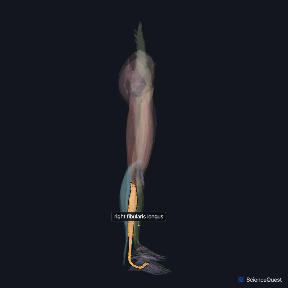
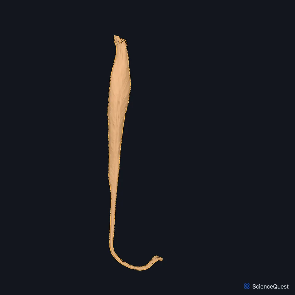
Open it in the Lower Limb Muscles Explorer See it alone in 3D
Where is the fibularis longus?
The lateral compartment is a narrow space walled by the crural fascia outside, the fibula inside and the anterior and posterior intermuscular septa. It holds the two fibular muscles, the common and superficial fibular nerves and branches of the anterior tibial and fibular arteries. (StatPearls, Leg Lateral Compartment)
It arises from the head and upper two-thirds of the lateral surface of the fibula and from the intermuscular septa. The fibularis brevis lies under it. (StatPearls, Leg Lateral Compartment; Gray’s Anatomy (1918), Lateral Crural Muscles)
What path does its tendon take?
Halfway down the compartment its fibers give way to a tendon, which runs behind the lateral malleolus in one synovial sheath with the fibularis brevis tendon, lying behind it. The superior fibular retinaculum holds the sheath in its groove. (StatPearls, Leg Lateral Compartment)
On the side of the calcaneus it runs below the fibular trochlea (peroneal tubercle), then makes a sharp turn at the cuboid and enters the sole through the cuboid tunnel. It inserts on the base of the first metatarsal and the medial cuneiform. (StatPearls, Leg Lateral Compartment and Calf Peroneus Longus Muscle; Gray’s Anatomy (1918), Calcaneus, p. 263)
The long plantar ligament turns the groove under the cuboid into a canal for the tendon. At the cuboid the tendon may carry an os peroneum, a fibrocartilaginous nodule that articulates with that bone. (Gray’s Anatomy (1918), Lateral Crural Muscles; StatPearls, Leg Lateral Compartment)
What does the fibularis longus do?
With the fibularis brevis it everts the foot and plantarflexes the ankle. The two muscles resist inversion, so an inversion ankle sprain can injure them. (StatPearls, Leg Lateral Compartment)
Its tendon crosses the transverse and lateral arches on its way to the first metatarsal. Along that course it helps hold both arches, and it lowers the first metatarsal as the foot pronates. (StatPearls, Leg Lateral Compartment)
With the tibialis anterior it forms a sling that StatPearls says lifts the arches. It also acts as a tie beam for the transverse arch. (StatPearls, Arches of the Foot)
On one leg, your weight tends to throw the leg medially, and the fibularis longus counters it by pulling on the lateral side of the leg. (Gray’s Anatomy (1918), Lateral Crural Muscles)
What supplies the fibularis longus?
The superficial fibular nerve supplies it. Its parent, the common fibular nerve, wraps around the neck of the fibula and passes between the attachments of the muscle, and in most people it divides there, in the fibular tunnel. (StatPearls, Calf Peroneus Longus Muscle and Leg Lateral Compartment)
The anterior tibial and fibular arteries supply it. (StatPearls, Calf Peroneus Longus Muscle)
Does the fibularis longus vary?
Its insertion varies. In one series of 30 cadaver specimens all reached the base of the first metatarsal and 86.6% the medial cuneiform, while some also reached the base of the second, fourth or fifth metatarsal. (StatPearls, Calf Peroneus Longus Muscle)
In rare cases the muscle has two bellies, each with its own tendon. (StatPearls, Leg Lateral Compartment)
Where the sources differ
Sources differ on its origin from the tibia: the StatPearls lateral compartment article lists the lateral tibial condyle among its origins, while Gray’s 1918 says only that a few fibers sometimes arise there. (StatPearls, Leg Lateral Compartment; Gray’s Anatomy (1918), Lateral Crural Muscles)
The StatPearls articles disagree on the rank of its actions: the fibularis longus article names plantarflexion and eversion as its primary functions, while the lateral compartment article makes eversion primary and plantarflexion secondary. (StatPearls, Calf Peroneus Longus Muscle and Leg Lateral Compartment)
| Feature | Detail and source |
|---|---|
| Origin | Head and upper two-thirds of the lateral fibula; intermuscular septa (Gray’s Anatomy (1918), Lateral Crural Muscles) |
| Insertion | Base of the first metatarsal and the medial cuneiform (StatPearls, Calf Peroneus Longus Muscle) |
| Action | Everts the foot; plantarflexes the ankle (StatPearls, Calf Peroneus Longus Muscle) |
| Nerve | Superficial fibular (superficial peroneal) nerve (StatPearls, Calf Peroneus Longus Muscle) |
Touches in this model
Measured on the BodyParts3D meshes: surfaces within 2 mm of the fibularis longus, nearest first, 12 of 19.
- Long head of right biceps femoris in contact
- Oblique head of right adductor hallucis in contact
- Right extensor digitorum longus in contact
- Right fibularis brevis in contact
- Right flexor hallucis longus in contact
- Right soleus in contact
- Right tibialis posterior in contact
- Short head of right biceps femoris in contact
- Right calcaneus in contact
- Right cuboid bone in contact
- Right fibula in contact
- Right first metatarsal bone in contact
Sources
Sources read
- StatPearls, Anatomy, Bony Pelvis and Lower Limb: Calf Peroneus Longus Muscle. Bradley Lezak; Matthew A. Varacallo. Last update . CC BY-NC-ND 4.0. Read .
- StatPearls, Anatomy, Bony Pelvis and Lower Limb: Leg Lateral Compartment. Irfan A. Khan; Navid Mahabadi; Anthony D’Abarno; Matthew A. Varacallo. Last update . CC BY-NC-ND 4.0. Read .
- Anatomy of the Human Body (Gray’s Anatomy), 20th US edition, 1918, Myology, The Muscles and Fasciae of the Leg, 3. The Lateral Crural Muscles, pages 486 to 488; Internet Archive scan of the 1918 Lea and Febiger printing, terminology of 1918. Henry Gray; edited by Warren H. Lewis. Public domain. Read .
- Gray’s Anatomy of the Human Body, 20th US edition (1918), edited by Warren H. Lewis, Osteology, The Calcaneus (pp. 263 to 266). Henry Gray; Warren H. Lewis (editor). Public domain. Read .
- StatPearls, Anatomy, Bony Pelvis and Lower Limb: Arches of the Foot. Hiteshkumar M. Chauhan; Muhammad Taqi. Last update . CC BY-NC-ND 4.0. Read .
The values are facts and free to use; this page’s selection and presentation are © 2026 ScienceQuest.