Long plantar ligament
The long plantar ligament is a ligament of the sole that attaches to the calcaneus and helps support the arch of the foot. Gray’s 1918 calls it the longest of all the ligaments of the tarsus. (StatPearls, Calcaneus; Gray’s Anatomy (1918), The Long Plantar Ligament)
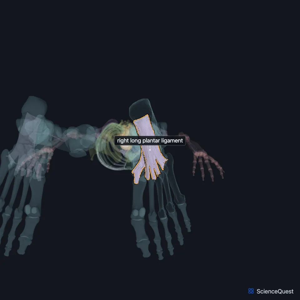
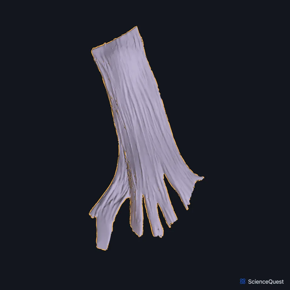
Where is the long plantar ligament?
Behind, it attaches to the plantar surface of the calcaneus in front of the tuberosity. In front it attaches to the tuberosity on the plantar surface of the cuboid, and its superficial fibers continue to the bases of the second, third and fourth metatarsals. (Gray’s Anatomy (1918), The Long Plantar Ligament)
On the bones themselves, Gray’s 1918 places it on a rough area of the calcaneus in front of the processes of the tuberosity, and on a prominent ridge under the cuboid, behind the groove for the fibularis longus tendon. (Gray’s Anatomy (1918), Calcaneus, p. 263, The Cuboid Bone and Lateral Crural Muscles)
Gray’s 1918 counts it among the five ligaments that join the calcaneus to the cuboid, and gives two older names, the long calcaneocuboid ligament and the superficial long plantar ligament. (Gray’s Anatomy (1918), The Long Plantar Ligament)
What lies deep to the long plantar ligament?
The plantar calcaneocuboid ligament, also called the short plantar ligament, lies nearer the bones, with a little areolar tissue between the two. It is short, wide and very strong, and runs from the front of the underside of the calcaneus to the cuboid behind the fibular groove. (Gray’s Anatomy (1918), The Long Plantar Ligament)
What does the long plantar ligament do?
It turns the groove on the underside of the cuboid into a canal for the tendon of the fibularis longus. StatPearls calls this fibro-osseous passage the cuboid tunnel. (Gray’s Anatomy (1918), The Long Plantar Ligament and Lateral Crural Muscles; StatPearls, Calf Peroneus Longus Muscle)
StatPearls lists it among the intersegmental ties that stabilize the lateral longitudinal arch. Gray’s 1918 says the long plantar and plantar calcaneocuboid ligaments, with the extensor tendons and the short muscles of the little toe, preserve that arch. (StatPearls, Arches of the Foot; Gray’s Anatomy (1918), Arches of the Foot)
In standing, the plantar ligaments and the plantar aponeurosis give the arches passive support. In walking, the muscles take over more of that work. (StatPearls, Arches of the Foot)
What does the long plantar ligament connect to?
The lateral arch it helps hold is made of the calcaneus, the cuboid and the fourth and fifth metatarsals, and its chief joint is the calcaneocuboid joint. StatPearls names the cuboid as the keystone of the arch and that joint as its most vulnerable region. (Gray’s Anatomy (1918), Arches of the Foot; StatPearls, Arches of the Foot)
The opponens digiti minimi of the foot, when present, arises from it and from the cuboid. (StatPearls, Foot and Ankle)
Injury to the spring ligament or to the long and short plantar ligaments is one cause of flatfoot, pes planus, along with posterior tibial tendon dysfunction and midfoot trauma. (StatPearls, Arches of the Foot)
| Feature | Detail and source |
|---|---|
| Attaches to | Plantar calcaneus; plantar tuberosity of the cuboid; bases of metatarsals 2 to 4 (Gray’s Anatomy (1918), The Long Plantar Ligament) |
| Joint | Calcaneocuboid joint (Gray’s Anatomy (1918), The Long Plantar Ligament) |
| Arch | One of the ties of the lateral longitudinal arch (StatPearls, Arches of the Foot) |
Touches in this model
Measured on the BodyParts3D meshes: surfaces within 2 mm of the long plantar ligament, nearest first, 12 of 22.
- Right calcaneus in contact
- Right first metatarsal bone in contact
- Right medial cuneiform bone in contact
- Right third metatarsal bone in contact
- Abductor digiti minimi of right foot in contact
- Flexor digiti minimi brevis of right foot in contact
- Lateral head of right flexor hallucis brevis in contact
- Medial head of right flexor hallucis brevis in contact
- Oblique head of right adductor hallucis in contact
- Opponens digiti minimi of right foot in contact
- Right abductor hallucis in contact
- Right fibularis longus in contact
Sources
Sources read
- StatPearls, Anatomy, Bony Pelvis and Lower Limb: Calcaneus. Marco Gupton; Meltem Özdemir; Robert R. Terreberry. Last update . CC BY-NC-ND 4.0. Read .
- Anatomy of the Human Body (Gray’s Anatomy), 20th US edition, 1918, Syndesmology, Intertarsal Articulations, Calcaneocuboid Articulation, the Long Plantar and Plantar Calcaneocuboid Ligaments, page 354; Internet Archive scan of the 1918 Lea and Febiger printing, terminology of 1918. Henry Gray; edited by Warren H. Lewis. Public domain. Read .
- Gray’s Anatomy of the Human Body, 20th US edition (1918), edited by Warren H. Lewis, Osteology, The Calcaneus (pp. 263 to 266). Henry Gray; Warren H. Lewis (editor). Public domain. Read .
- Anatomy of the Human Body (Gray’s Anatomy), 20th US edition, 1918, Osteology, The Foot, The Cuboid Bone, page 269; Internet Archive scan of the 1918 Lea and Febiger printing, terminology of 1918. Henry Gray; edited by Warren H. Lewis. Public domain. Read .
- Anatomy of the Human Body (Gray’s Anatomy), 20th US edition, 1918, Myology, The Muscles and Fasciae of the Leg, 3. The Lateral Crural Muscles, pages 486 to 488; Internet Archive scan of the 1918 Lea and Febiger printing, terminology of 1918. Henry Gray; edited by Warren H. Lewis. Public domain. Read .
- StatPearls, Anatomy, Bony Pelvis and Lower Limb: Calf Peroneus Longus Muscle. Bradley Lezak; Matthew A. Varacallo. Last update . CC BY-NC-ND 4.0. Read .
- StatPearls, Anatomy, Bony Pelvis and Lower Limb: Arches of the Foot. Hiteshkumar M. Chauhan; Muhammad Taqi. Last update . CC BY-NC-ND 4.0. Read .
- Anatomy of the Human Body (Gray’s Anatomy), 20th US edition, 1918, Syndesmology, Arches of the Foot, page 360; Internet Archive scan of the 1918 Lea and Febiger printing, terminology of 1918. Henry Gray; edited by Warren H. Lewis. Public domain. Read .
- StatPearls, Anatomy, Bony Pelvis and Lower Limb: Foot and Ankle. Jennifer Ficke; Adegbenro O. Fakoya; Doug W. Byerly. Last update . CC BY-NC-ND 4.0. Read .
The values are facts and free to use; this page’s selection and presentation are © 2026 ScienceQuest.