Metatarsal bones
The five metatarsals are the bones of the forefoot, between the tarsal bones and the toes. They are numbered one to five from the big toe. (StatPearls, Metatarsal Bones; Gray’s Anatomy (1918), Metatarsus, p. 272)
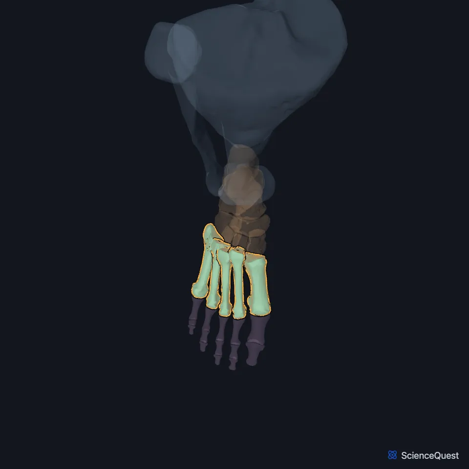
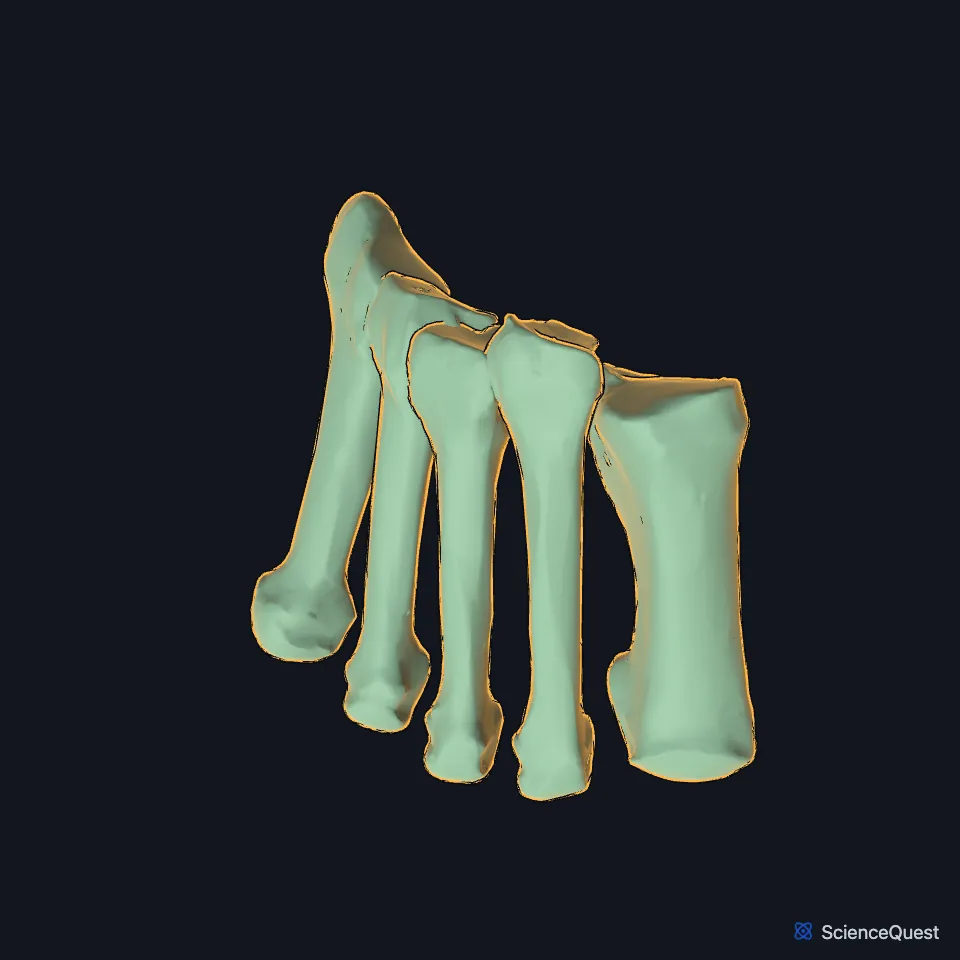
Open it in the Ankle and Foot Skeleton Explorer See it alone in 3D
Where are the metatarsals?
They connect the three cuneiforms and the cuboid behind to the toes in front. The base of each meets the tarsus at the tarsometatarsal, or Lisfranc, joint complex, and its head meets a proximal phalanx at a metatarsophalangeal joint. (StatPearls, Metatarsal Bones)
Which tarsal bones does each metatarsal meet?
Gray’s Anatomy (1918): the first meets the medial cuneiform, the second all three cuneiforms, the third the lateral cuneiform, the fourth the lateral cuneiform and cuboid, and the fifth the cuboid. (Gray’s Anatomy (1918), Metatarsus, p. 272)
Tendons from the leg end on them. Fibularis longus and tibialis anterior insert on the first, tibialis posterior on the second to fourth, and fibularis brevis and fibularis tertius on the fifth. (StatPearls, Metatarsal Bones)
How do the five metatarsals differ?
The first is the shortest and the thickest, with two grooved facets under its head for sesamoid bones. The second is the longest, its base set back into a recess between the three cuneiforms, and the fifth has a rough tuberosity on the lateral side of its base. (Gray’s Anatomy (1918), Metatarsus, p. 272; StatPearls, Metatarsal Bones)
What do the metatarsals do?
Their heads are a main weight-bearing area of the foot, and the first metatarsal carries about 30% to 50% of the load during walking. The first and fifth can move up and down to meet uneven ground, while the bases of the middle three are held fixed. (StatPearls, Metatarsal Bones)
They are part of all three arches. The first three are part of the medial longitudinal arch and the heads of the fourth and fifth part of the lateral arch, and their bases, with the cuboid and cuneiforms, form the transverse arch. (StatPearls, Metatarsal Bones)
Which metatarsals break most often?
Metatarsal fractures are among the commonest foot injuries. Adults, adolescents and older children break the fifth metatarsal most often, and children under 5 the first. Stress fractures favour the bases of the first to third and the necks of the second to fourth. (StatPearls, Metatarsal Fractures, Metatarsal Bones and Foot and Ankle)
Where the sources differ
StatPearls pairs the second metatarsal with the intermediate cuneiform, in the middle column of the tarsometatarsal joints. Gray’s Anatomy (1918) has the base of the second meeting all three cuneiforms. (StatPearls, Metatarsal Bones; Gray’s Anatomy (1918), Metatarsus, p. 272)
| Feature | Detail and source |
|---|---|
| Number | Five, numbered from the big toe (StatPearls, Metatarsal Bones) |
| Shortest and longest | First shortest, second longest (StatPearls, Metatarsal Bones; Gray’s Anatomy (1918), Metatarsus, p. 272) |
| Joints | Tarsometatarsal (Lisfranc) behind, metatarsophalangeal in front (StatPearls, Metatarsal Bones) |
Touches in this model
Measured on the BodyParts3D meshes: surfaces within 2 mm of the metatarsal bones, nearest first, 12 of 37.
- Proximal phalanx of right big toe in contact
- Proximal phalanx of right fourth toe in contact
- Proximal phalanx of right little toe in contact
- Proximal phalanx of right second toe in contact
- Proximal phalanx of right third toe in contact
- Right cuboid bone in contact
- Right lateral cuneiform bone in contact
- Right medial cuneiform bone in contact
- Abductor digiti minimi of right foot in contact
- First lumbrical of right foot in contact
- First plantar interosseous of right foot in contact
- Flexor digiti minimi brevis of right foot in contact
Sources
Sources read
- StatPearls, Anatomy, Bony Pelvis and Lower Limb: Metatarsal Bones. Bradley Lezak; James La Rose; Dustin H. Massel. Last update . CC BY-NC-ND 4.0. Read .
- Gray’s Anatomy of the Human Body, 20th US edition (1918), edited by Warren H. Lewis, Osteology, The Metatarsus (pp. 272 to 274); 1918 names: first, second and third cuneiform for medial, intermediate and lateral cuneiform. Henry Gray; Warren H. Lewis (editor). Public domain. Read .
- StatPearls, Metatarsal Fractures. Udit Agrawal; Vivek Tiwari. Last update . CC BY-NC-ND 4.0. Read .
- StatPearls, Anatomy, Bony Pelvis and Lower Limb: Foot and Ankle. Jennifer Ficke; Adegbenro O. Fakoya; Doug W. Byerly. Last update . CC BY-NC-ND 4.0. Read .
The values are facts and free to use; this page’s selection and presentation are © 2026 ScienceQuest.