Iliotibial tract
The iliotibial tract is a long band of thickened fascia lata running down the outer side of the thigh from the hip to the tibia. It is also called the iliotibial band, or Maissiat’s band. (StatPearls, Iliotibial Band (Tract); Gray’s Anatomy (1918), Anterior Femoral Muscles)
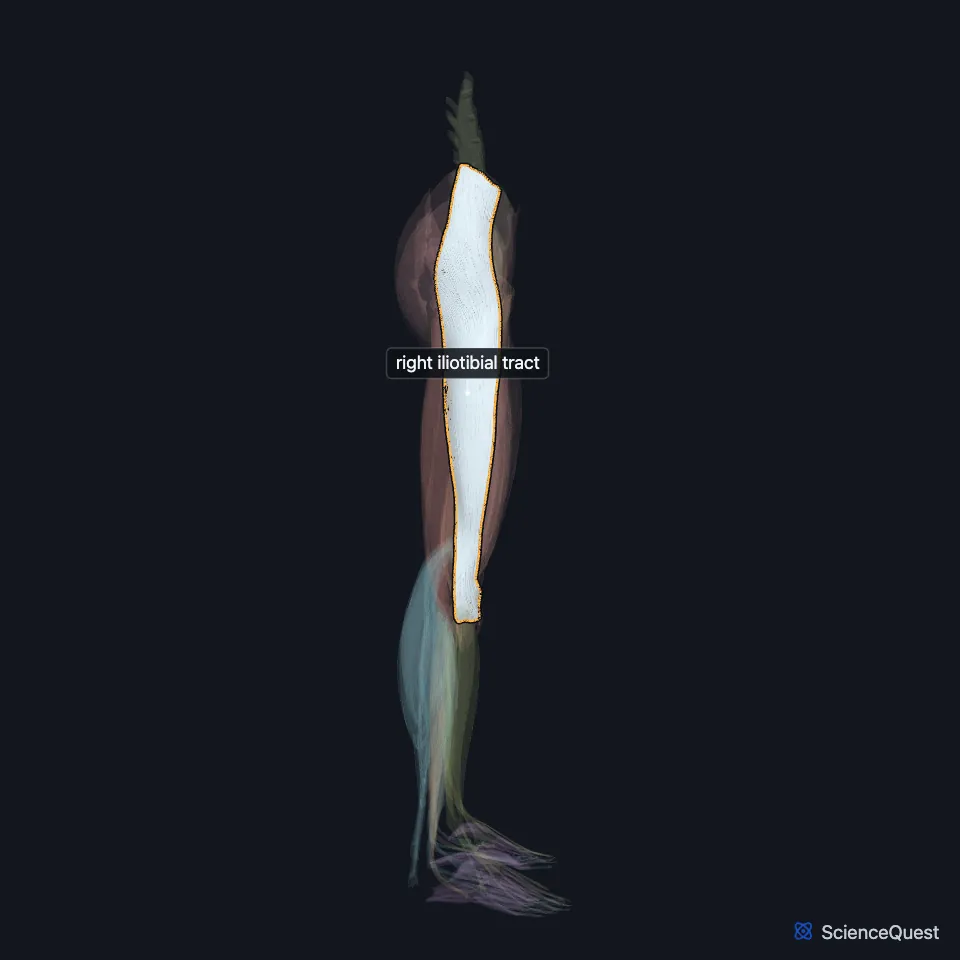
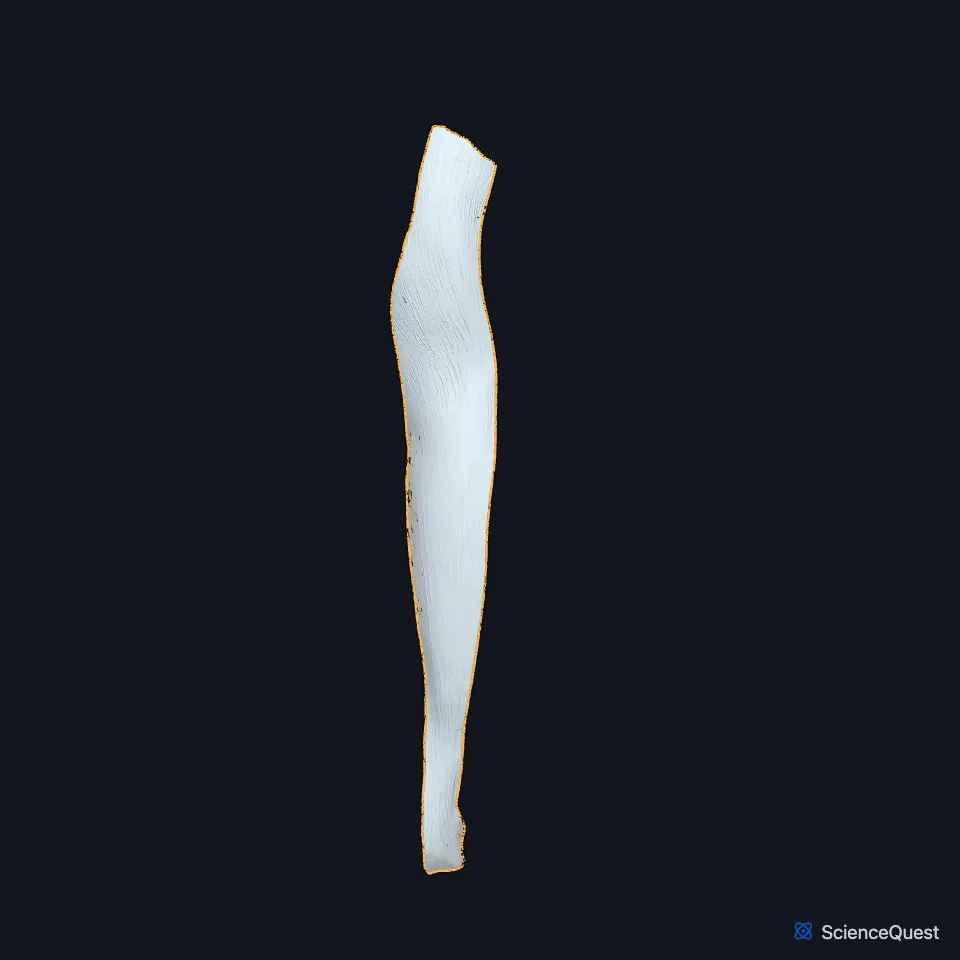
Open it in the Lower Limb Muscles Explorer See it alone in 3D
Where is the iliotibial tract?
It runs along the lateral thigh and ends on Gerdy’s tubercle, on the upper outer tibia. (StatPearls, Iliotibial Band (Tract))
Above the knee it attaches to the intermuscular septum and the supracondylar tubercle of the femur. Just above the lateral epicondyle, a layer of fat lies between it and the vastus lateralis. (StatPearls, Iliotibial Band (Tract))
How is the iliotibial tract formed?
Gray’s (1918) describes the fascia lata splitting into two layers around the tensor fasciae latae. Below the muscle the layers unite into a strong band, after receiving the muscle’s insertion. (Gray’s Anatomy (1918), Anterior Femoral Muscles)
The gluteus maximus also inserts into it. Its upper fibers and the superficial fibers of its lower part end in a tendinous sheet that crosses the greater trochanter into the band. (Gray’s Anatomy (1918), Muscles of the Gluteal Region; StatPearls, Iliotibial Band (Tract))
A 2013 study StatPearls cites describes three layers in the upper tract, superficial, intermediate and deep, which fuse near the greater trochanter. The deep layer blends with the hip joint capsule, as Gray’s (1918) also describes. (StatPearls, Iliotibial Band (Tract); Gray’s Anatomy (1918), Anterior Femoral Muscles)
What does the iliotibial tract do?
At the hip it helps extend, abduct and laterally rotate. At the knee its action depends on position: from full extension to about 20° to 30° of flexion it lies in front of the lateral femoral epicondyle and helps extend, and beyond that it lies behind the epicondyle and helps flex. (StatPearls, Iliotibial Band (Tract))
Gray’s (1918) says that through the band the gluteus maximus steadies the femur on the tibia in standing, and the tensor fasciae latae helps it hold the knee extended. (Gray’s Anatomy (1918), Muscles of the Gluteal Region)
Which nerves, vessels and septum go with the iliotibial tract?
It shares the nerves of the muscles that pull on it: the superior gluteal nerve through the tensor fasciae latae, and the inferior gluteal nerve through the gluteus maximus. (StatPearls, Iliotibial Band (Tract))
It shares the blood supply of the tensor fasciae latae: the ascending branch of the lateral circumflex femoral artery and the superior gluteal artery. (StatPearls, Iliotibial Band (Tract))
From the deep surface of the fascia lata, the lateral intermuscular septum runs to the linea aspera. It separates the vastus lateralis in front from the short head of the biceps femoris behind. (Gray’s Anatomy (1918), Anterior Femoral Muscles; StatPearls, Fascia Lata)
What problems involve the iliotibial tract?
Iliotibial band syndrome is an overuse injury of runners and cyclists that causes pain on the outer knee, worst at about 30° to 40° of knee flexion. Its cause is debated: friction over the lateral femoral epicondyle, compression of the fat beneath the band, or inflammation of its bursa. (StatPearls, Iliotibial Band (Tract) and Tensor Fasciae Latae Muscle)
In external snapping hip, the back part of the band, or the front tendon fibers of the gluteus maximus near their insertion, thicken. The thickened part of the band sits behind the greater trochanter with the hip extended and moves forward, with a snap, as the hip flexes, adducts or rotates medially. Clinicians examine the band with the Ober test. (StatPearls, Iliotibial Band (Tract))
| Feature | Detail and source |
|---|---|
| Formed from | Fascia lata, with fibers from the tensor fasciae latae and the gluteus maximus (StatPearls, Iliotibial Band (Tract)) |
| Lower attachment | Gerdy’s tubercle, on the lateral condyle of the tibia (StatPearls, Iliotibial Band (Tract); Gray’s Anatomy (1918), Anterior Femoral Muscles) |
| Function | Helps extend, abduct and laterally rotate the hip; at the knee, helps extend near full extension and helps flex past 20° to 30° (StatPearls, Iliotibial Band (Tract)) |
| Nerve | Superior gluteal nerve (through the tensor fasciae latae); inferior gluteal nerve (through the gluteus maximus) (StatPearls, Iliotibial Band (Tract)) |
Touches in this model
Measured on the BodyParts3D meshes: surfaces within 2 mm of the iliotibial tract, nearest first, 12 of 16.
- Right extensor digitorum longus in contact
- Right gluteus maximus in contact
- Right gluteus medius in contact
- Right gluteus minimus in contact
- Right rectus femoris in contact
- Right tensor fasciae latae in contact
- Right tibialis anterior in contact
- Right vastus intermedius in contact
- Right vastus lateralis in contact
- Right femur in contact
- Right hip bone in contact
- Right tibia in contact
Sources
Sources read
- StatPearls, Anatomy, Bony Pelvis and Lower Limb, Iliotibial Band (Tract). Scott Hyland; Steven B. Graefe; Matthew A. Varacallo. Last update . CC BY-NC-ND 4.0. Read .
- Anatomy of the Human Body (Gray’s Anatomy), 20th US edition, 1918, Myology, The Muscles and Fasciae of the Thigh, 1. The Anterior Femoral Muscles, pages 467 to 471; Internet Archive scan of the 1918 Lea and Febiger printing, terminology of 1918. Henry Gray; edited by Warren H. Lewis. Public domain. Read .
- Anatomy of the Human Body (Gray’s Anatomy), 20th US edition, 1918, Myology, The Muscles and Fasciae of the Thigh, 3. The Muscles of the Gluteal Region, pages 474 to 478; Internet Archive scan of the 1918 Lea and Febiger printing, terminology of 1918. Henry Gray; edited by Warren H. Lewis. Public domain. Read .
- StatPearls, Anatomy, Bony Pelvis and Lower Limb: Fascia Lata. Tucker Peabody; Bruno Bordoni. Last update . CC BY-NC-ND 4.0. Read .
- StatPearls, Anatomy, Bony Pelvis and Lower Limb: Tensor Fasciae Latae Muscle. Amy P. Trammell; Ahmed Nahian; Holly Pilson. Last update . CC BY-NC-ND 4.0. Read .
The values are facts and free to use; this page’s selection and presentation are © 2026 ScienceQuest.