Tensor fasciae latae
The tensor fasciae latae is a small muscle at the front and outer side of the upper thigh. It lies between the superficial and deep layers of the iliotibial tract, through which it acts on the tibia. (StatPearls, Tensor Fasciae Latae Muscle)
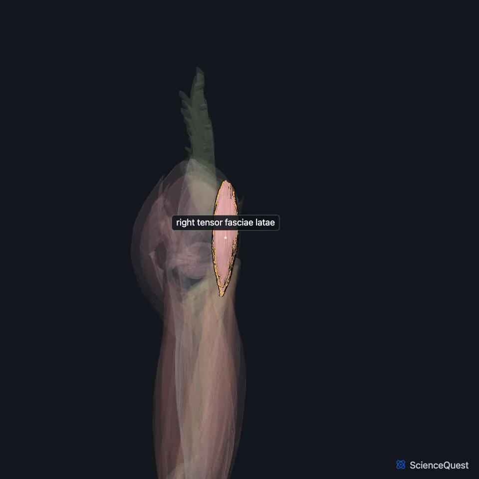
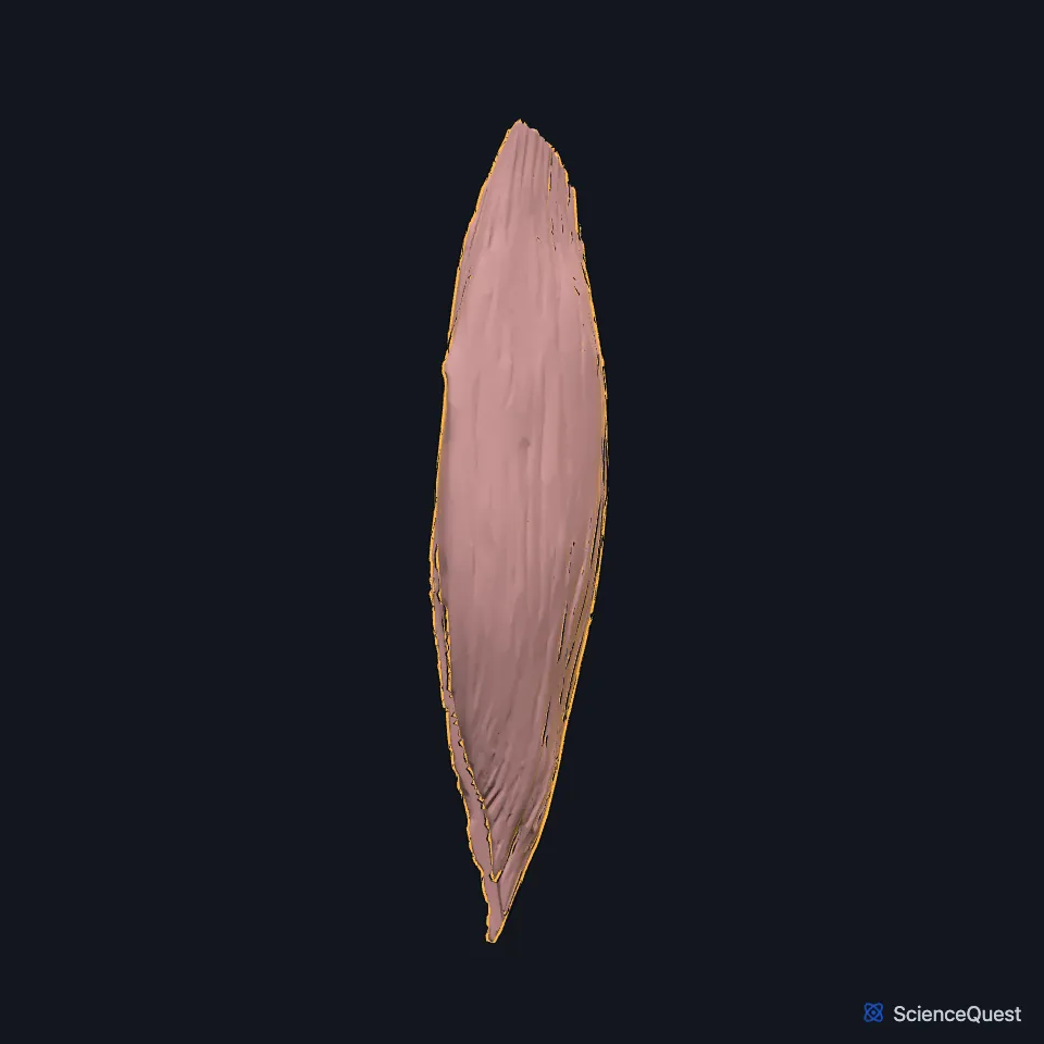
Open it in the Lower Limb Muscles Explorer See it alone in 3D
Where is the tensor fasciae latae?
It arises from the anterior superior iliac spine and the front of the iliac crest. It then runs down the front and outer side of the thigh, superficial to the greater trochanter. (StatPearls, Tensor Fasciae Latae Muscle)
Gray’s (1918) places its origin between the gluteus medius and the sartorius, and notes that it also arises from the deep surface of the fascia lata. (Gray’s Anatomy (1918), Muscles of the Gluteal Region)
Where does the tensor fasciae latae insert?
It inserts between the two layers of the iliotibial tract, at about the junction of the upper and middle thirds of the thigh. The tract then runs down the outer thigh to Gerdy’s tubercle on the lateral condyle of the tibia. (Gray’s Anatomy (1918), Muscles of the Gluteal Region; StatPearls, Tensor Fasciae Latae Muscle)
What does the tensor fasciae latae do?
With the gluteus medius and gluteus minimus it abducts the hip and rotates it medially. Through the iliotibial tract it also works with the gluteus maximus to abduct the hip, and it helps the rectus femoris flex the hip. (StatPearls, Tensor Fasciae Latae Muscle)
In walking, it pulls the ilium down on the weight-bearing side. That raises the other side of the pelvis, so the swinging leg clears the ground. (StatPearls, Tensor Fasciae Latae Muscle)
Through the iliotibial tract it steadies the knee in full extension. It helps flex the knee only once the knee is bent past about 30°. (StatPearls, Tensor Fasciae Latae Muscle)
It can tighten after long periods held short, such as sitting. A shortened tensor fasciae latae can tilt the pelvis forward or rotate the femur medially. (StatPearls, Tensor Fasciae Latae Muscle)
Which nerve and vessels supply the tensor fasciae latae?
The superior gluteal nerve (L4, L5 and S1) supplies it, the same nerve as the gluteus medius and gluteus minimus. The nerve leaves the pelvis through the greater sciatic foramen above the piriformis. (StatPearls, Tensor Fasciae Latae Muscle and Superior Gluteal Nerve)
The deep branch of the superior gluteal artery supplies it, running between the gluteus minimus and gluteus medius on the way. (StatPearls, Tensor Fasciae Latae Muscle)
Surgeons use it as a landmark. The anterior approach to the hip passes between the sartorius, supplied by the femoral nerve, and the tensor fasciae latae, supplied by the superior gluteal nerve. (StatPearls, Tensor Fasciae Latae Muscle)
How does the tensor fasciae latae vary?
The length of its belly varies. In most people it ends before the greater trochanter, but in about one third it reaches past it, and a surgeon making a lateral approach to the upper femur must then split its fibers. (StatPearls, Tensor Fasciae Latae Muscle)
Where the sources differ
Sources disagree on its actions. The StatPearls muscle article has it helping flex the hip and, past 30°, flex the knee; the StatPearls superior gluteal nerve article has it helping the gluteus maximus extend the hip and knee, and Gray’s (1918) has it helping hold the knee extended. (StatPearls, Tensor Fasciae Latae Muscle and Superior Gluteal Nerve; Gray’s Anatomy (1918), Muscles of the Gluteal Region)
Sources disagree on its nerve. The StatPearls tensor fasciae latae article names only the superior gluteal nerve, while the StatPearls fascia lata article says the inferior gluteal nerve also supplies the muscle. (StatPearls, Tensor Fasciae Latae Muscle and Fascia Lata)
| Feature | Detail and source |
|---|---|
| Origin | Anterior superior iliac spine and the front of the iliac crest (StatPearls, Tensor Fasciae Latae Muscle) |
| Insertion | Between the two layers of the iliotibial tract, which attaches to Gerdy’s tubercle of the tibia (Gray’s Anatomy (1918), Muscles of the Gluteal Region; StatPearls, Tensor Fasciae Latae Muscle) |
| Action | Abducts and medially rotates the hip; helps flex the hip; steadies the extended knee (StatPearls, Tensor Fasciae Latae Muscle) |
| Nerve | Superior gluteal nerve (L4, L5, S1) (StatPearls, Tensor Fasciae Latae Muscle) |
Touches in this model
Measured on the BodyParts3D meshes: surfaces within 2 mm of the tensor fasciae latae, nearest first.
- Right iliotibial tract in contact
- Right rectus femoris in contact
- Right sartorius in contact
- Right hip bone in contact
- Skin in contact
- Right iliacus 1.9 mm away
Sources
Sources read
- StatPearls, Anatomy, Bony Pelvis and Lower Limb: Tensor Fasciae Latae Muscle. Amy P. Trammell; Ahmed Nahian; Holly Pilson. Last update . CC BY-NC-ND 4.0. Read .
- Anatomy of the Human Body (Gray’s Anatomy), 20th US edition, 1918, Myology, The Muscles and Fasciae of the Thigh, 3. The Muscles of the Gluteal Region, pages 474 to 478; Internet Archive scan of the 1918 Lea and Febiger printing, terminology of 1918. Henry Gray; edited by Warren H. Lewis. Public domain. Read .
- StatPearls, Anatomy, Abdomen and Pelvis: Superior Gluteal Nerve. Kirsten Lung; Forshing Lui. Last update . CC BY-NC-ND 4.0. Read .
- StatPearls, Anatomy, Bony Pelvis and Lower Limb: Fascia Lata. Tucker Peabody; Bruno Bordoni. Last update . CC BY-NC-ND 4.0. Read .
The values are facts and free to use; this page’s selection and presentation are © 2026 ScienceQuest.