Vastus intermedius
The vastus intermedius is one of the four muscles of the quadriceps femoris. It arises from the front and outer surfaces of the femur and forms the deep part of the quadriceps tendon. (StatPearls, Anterior Thigh Muscles; Gray’s Anatomy (1918), Anterior Femoral Muscles)
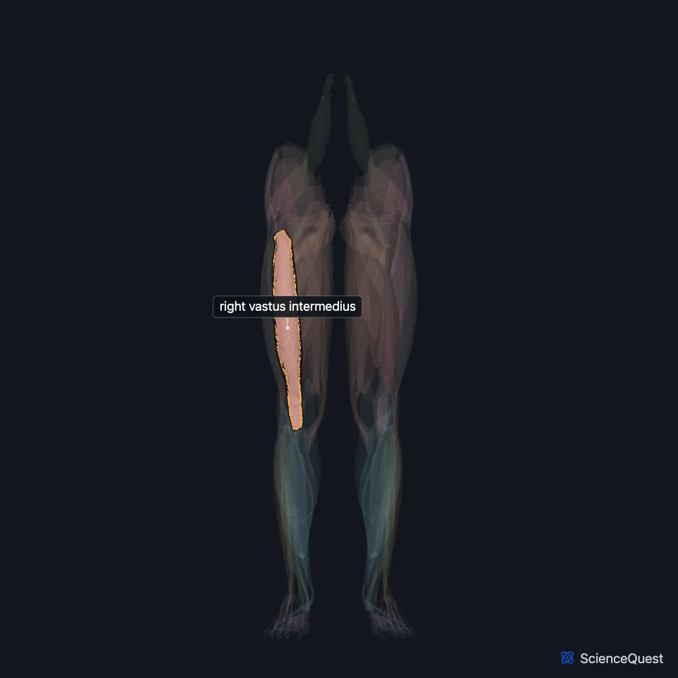
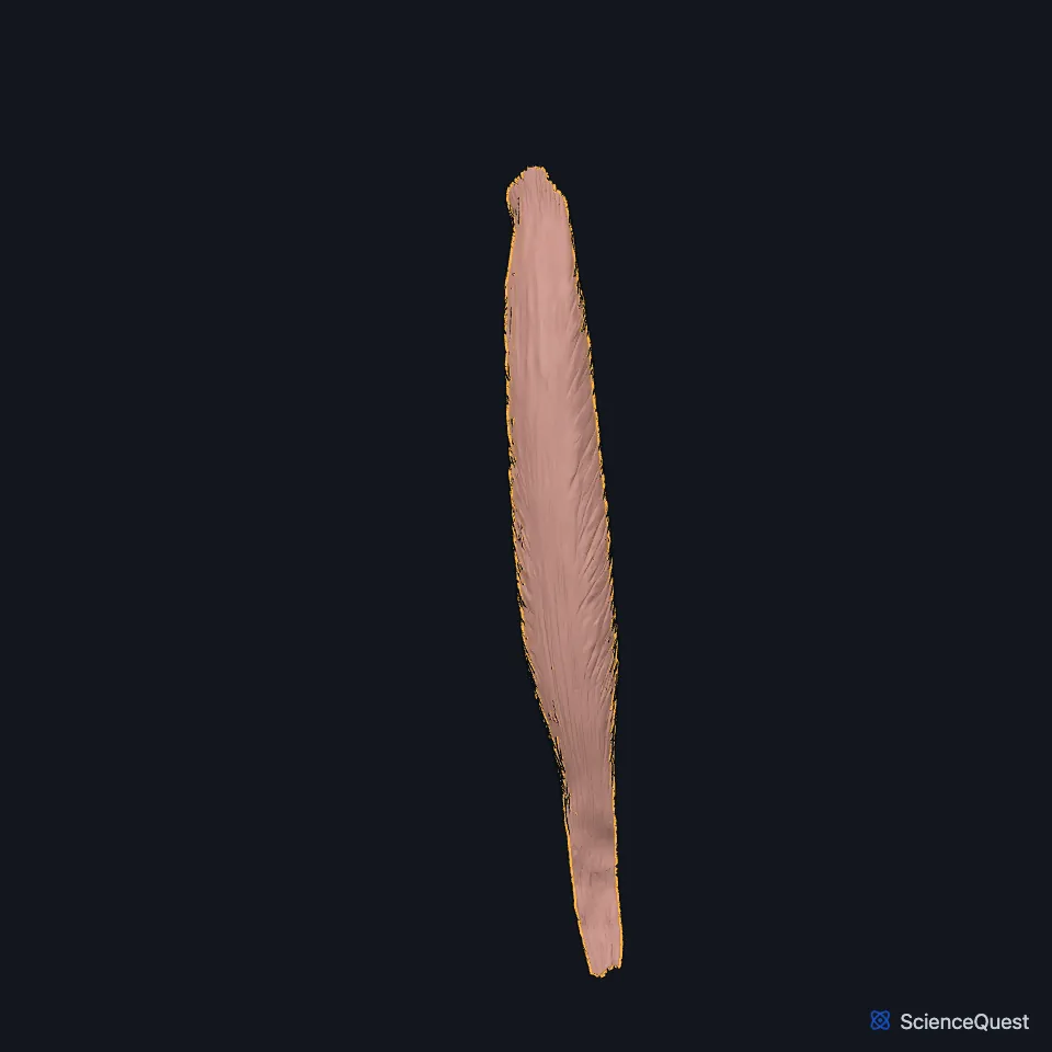
Open it in the Lower Limb Muscles Explorer See it alone in 3D
Where is the vastus intermedius?
It arises from the front and lateral surfaces of the upper shaft of the femur and from the lower part of the lateral intermuscular septum. (Gray’s Anatomy (1918), Anterior Femoral Muscles; StatPearls, Thigh Quadriceps Muscle)
It is hard to separate from the vastus medialis. With the rectus femoris lifted away, a narrow gap between the two runs up from the medial border of the patella, but higher up the muscles are often continuous. (Gray’s Anatomy (1918), Anterior Femoral Muscles)
How does the vastus intermedius reach the patella?
With the other three parts it joins the quadriceps tendon, which attaches to the patella and continues to the tuberosity of the tibia. (StatPearls, Anterior Thigh Muscles)
Some of its bundles insert into the upper recess of the suprapatellar bursa and make up the articular muscle of the knee. (StatPearls, Thigh Quadriceps Muscle)
A more recently described muscle, the tensor of the vastus intermedius, starts from the lower front of the greater trochanter and joins a flat aponeurosis between the vastus lateralis and the vastus intermedius. (StatPearls, Thigh Quadriceps Muscle)
What does the vastus intermedius do?
It extends the knee, together with the vastus lateralis, vastus medialis and rectus femoris. (StatPearls, Thigh Quadriceps Muscle; Gray’s Anatomy (1918), Anterior Femoral Muscles)
The three vasti extend the knee and steady the patella. Of the four parts of the quadriceps, only the rectus femoris also flexes the hip. (StatPearls, Anterior Thigh Muscles)
In sport, the vastus lateralis and vastus intermedius are the vasti most often hurt by direct blows, being the most exposed. A contusion that is not treated properly can lead to myositis ossificans. (StatPearls, Thigh Quadriceps Muscle)
Which nerve supplies the vastus intermedius?
The femoral nerve supplies it through the nerve to the vastus intermedius, one of the four motor branches of the nerve’s posterior division. That branch also supplies the knee joint. (StatPearls, Thigh Femoral Nerve)
Its nerve fibers come from the second, third and fourth lumbar nerves (L2 to L4), which reach the quadriceps through the femoral nerve. (Gray’s Anatomy (1918), Anterior Femoral Muscles; StatPearls, Anterior Thigh Muscles)
Where the sources differ
Sources differ on how far down the femur it arises. Gray’s (1918) gives the upper two thirds of the shaft; the StatPearls quadriceps article gives the upper three fourths and adds the lateral lip of the linea aspera. (Gray’s Anatomy (1918), Anterior Femoral Muscles; StatPearls, Thigh Quadriceps Muscle)
Sources differ on the articular muscle of the knee. The StatPearls quadriceps article describes it as bundles of the vastus intermedius; Gray’s (1918) calls it a small muscle usually distinct from the vastus intermedius and only occasionally blended with it. (StatPearls, Thigh Quadriceps Muscle; Gray’s Anatomy (1918), Anterior Femoral Muscles)
Two StatPearls articles place its patellar attachment differently: the anterior thigh article at the lateral border of the patella, the quadriceps article at the base. (StatPearls, Anterior Thigh Muscles and Thigh Quadriceps Muscle)
| Feature | Detail and source |
|---|---|
| Origin | Front and lateral surfaces of the upper femoral shaft; lower lateral intermuscular septum (Gray’s Anatomy (1918), Anterior Femoral Muscles; StatPearls, Anterior Thigh Muscles) |
| Insertion | Deep part of the quadriceps tendon, to the patella (StatPearls, Anterior Thigh Muscles; Gray’s Anatomy (1918), Anterior Femoral Muscles) |
| Action | Extends the knee (StatPearls, Thigh Quadriceps Muscle) |
| Nerve | Femoral nerve (L2 to L4), posterior division (StatPearls, Thigh Femoral Nerve and Anterior Thigh Muscles) |
Touches in this model
Measured on the BodyParts3D meshes: surfaces within 2 mm of the vastus intermedius, nearest first.
- Right iliotibial tract in contact
- Right rectus femoris in contact
- Right vastus lateralis in contact
- Right vastus medialis in contact
- Right femur in contact
- Right patella in contact
- Right tibia in contact
- Skin in contact
Sources
Sources read
- StatPearls, Anatomy, Bony Pelvis and Lower Limb: Anterior Thigh Muscles. Ayesha Khan; Abdul Arain. Last update . CC BY-NC-ND 4.0. Read .
- Anatomy of the Human Body (Gray’s Anatomy), 20th US edition, 1918, Myology, The Muscles and Fasciae of the Thigh, 1. The Anterior Femoral Muscles, pages 467 to 471; Internet Archive scan of the 1918 Lea and Febiger printing, terminology of 1918. Henry Gray; edited by Warren H. Lewis. Public domain. Read .
- StatPearls, Anatomy, Bony Pelvis and Lower Limb: Thigh Quadriceps Muscle. Bruno Bordoni; Matthew A. Varacallo. Last update . CC BY-NC-ND 4.0. Read .
- StatPearls, Anatomy, Bony Pelvis and Lower Limb: Thigh Femoral Nerve. Nader A. Refai; Asa C. Black; Prasanna Tadi. Last update . CC BY-NC-ND 4.0. Read .
The values are facts and free to use; this page’s selection and presentation are © 2026 ScienceQuest.