Interosseous membrane of leg
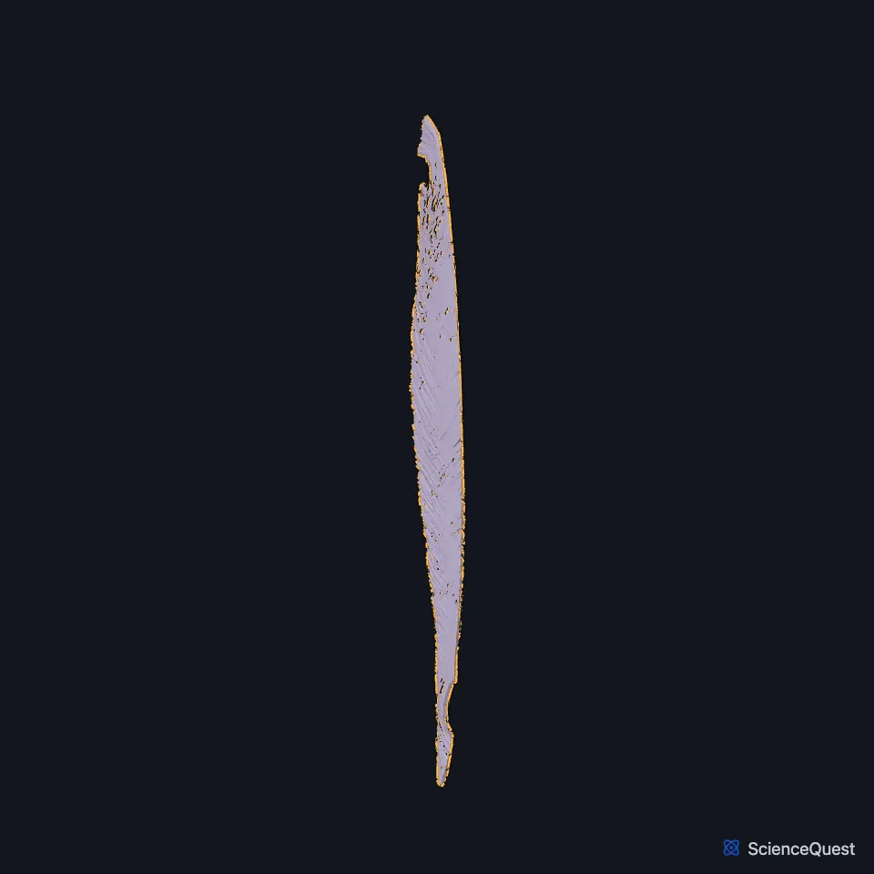
The interosseous membrane of the leg is a thin sheet of oblique fibers stretched between the tibia and the fibula. It separates the muscles on the front of the leg from those on the back. (Gray’s Anatomy (1918), Tibiofibular joints, p. 347)
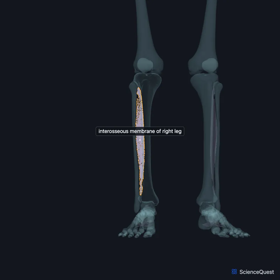
Where is the interosseous membrane of the leg?
It attaches to the interosseous crests of the two bones. On the fibula, StatPearls describes this attachment as a ridge on the medial surface, the interosseous border, and the joint it forms as a syndesmosis with very little movement. (Gray’s Anatomy (1918), Tibiofibular joints, p. 347; StatPearls, Fibula)
With the anterior and posterior intermuscular septa and the crural fascia, it divides the leg into its anterior, lateral and posterior compartments. (StatPearls, Leg Posterior Compartment)
How is the interosseous membrane built?
Most of its fibers run downward and laterally, and a few run the other way. It is broader above than below, and Gray’s 1918 also names it the middle tibiofibular ligament. (Gray’s Anatomy (1918), Tibiofibular joints, p. 347)
Its upper edge stops short of the upper tibiofibular joint and leaves a large oval opening. The anterior tibial artery passes forward through it, between the two heads of the tibialis posterior, to the front of the leg. (Gray’s Anatomy (1918), Tibiofibular joints, p. 347; StatPearls, Tibial Nerve)
Lower down it has an opening for what Gray’s 1918 calls the anterior peroneal vessels and many small holes for small vessels. At its lower end it continues into the interosseous ligament of the distal tibiofibular syndesmosis. (Gray’s Anatomy (1918), Tibiofibular joints, p. 347; StatPearls, Distal Tibiofibular Joint)
What does the interosseous membrane of the leg do?
It gives origin to muscles on its front: the tibialis anterior, extensor hallucis longus, extensor digitorum longus and fibularis tertius all take part of their origin from it. (StatPearls, Leg Anterior Compartment)
From its back arise the tibialis posterior and part of the flexor hallucis longus. (StatPearls, Leg Posterior Compartment and Tibial Nerve)
The interosseous ligament that continues it at the ankle works as a spring-like stabilizer. It limits separation of the tibia and fibula in ankle dorsiflexion and at heel strike. (StatPearls, Distal Tibiofibular Joint)
What lies on each side of the membrane?
In front lie the tibialis anterior, the long extensors, the anterior tibial vessels and the deep fibular nerve. Behind lie the tibialis posterior and the flexor hallucis longus. (Gray’s Anatomy (1918), Tibiofibular joints, p. 347; StatPearls, Arches of the Foot)
StatPearls counts the membrane, with the interosseous ligament and the anterior and posterior inferior tibiofibular ligaments, as part of the distal tibiofibular syndesmosis above the ankle. Injury to these structures is called a high ankle sprain. (StatPearls, Foot and Ankle)
| Feature | Detail and source |
|---|---|
| Between | Interosseous crests of the tibia and fibula (Gray’s Anatomy (1918), Tibiofibular joints, p. 347) |
| Joint type | Syndesmosis, with very little movement (StatPearls, Fibula) |
| Main opening | Upper oval opening for the anterior tibial vessels (Gray’s Anatomy (1918), Tibiofibular joints, p. 347) |
Touches in this model
Measured on the BodyParts3D meshes: surfaces within 2 mm of the interosseous membrane of leg, nearest first.
- Right tibia in contact
- Right extensor digitorum longus in contact
- Right extensor hallucis longus in contact
- Right fibularis tertius in contact
- Right flexor hallucis longus in contact
- Right tibialis anterior in contact
- Right tibialis posterior in contact
- Skin in contact
- Right fibula 0.7 mm away
Sources
Sources read
- Gray’s Anatomy of the Human Body, 20th US edition (1918), edited by Warren H. Lewis, Syndesmology, Tibiofibular Articulation and Tibiofibular Syndesmosis (pp. 347 to 349). Henry Gray; Warren H. Lewis (editor). Public domain. Read .
- StatPearls, Anatomy, Bony Pelvis and Lower Limb: Fibula. Marco Gupton; Akul Munjal; Michael Kang. Last update . CC BY-NC-ND 4.0. Read .
- StatPearls, Anatomy, Bony Pelvis and Lower Limb: Leg Posterior Compartment. Rami Abuqubo; Steven B. Graefe; James La Rose. Last update . CC BY-NC-ND 4.0. Read .
- StatPearls, Anatomy, Bony Pelvis and Lower Limb: Tibial Nerve. Sohil S. Desai; James La Rose. Last update . CC BY-NC-ND 4.0. Read .
- StatPearls, Anatomy, Bony Pelvis and Lower Limb: Distal Tibiofibular Joint (Tibiofibular Syndesmosis). Bradley Lezak; Matthew A. Varacallo. Last update . CC BY-NC-ND 4.0. Read .
- StatPearls, Anatomy, Bony Pelvis and Lower Limb: Leg Anterior Compartment. Bradley Lezak; Marjorie V. Launico. Last update . CC BY-NC-ND 4.0. Read .
- StatPearls, Anatomy, Bony Pelvis and Lower Limb: Arches of the Foot. Hiteshkumar M. Chauhan; Muhammad Taqi. Last update . CC BY-NC-ND 4.0. Read .
- StatPearls, Anatomy, Bony Pelvis and Lower Limb: Foot and Ankle. Jennifer Ficke; Adegbenro O. Fakoya; Doug W. Byerly. Last update . CC BY-NC-ND 4.0. Read .
The values are facts and free to use; this page’s selection and presentation are © 2026 ScienceQuest.