Putamen
The putamen is one of the basal nuclei deep in the cerebrum. With the caudate nucleus it forms the striatum, and with the globus pallidus the lentiform nucleus. (OpenStax A&P 2e, 13.2; StatPearls, Putamen)
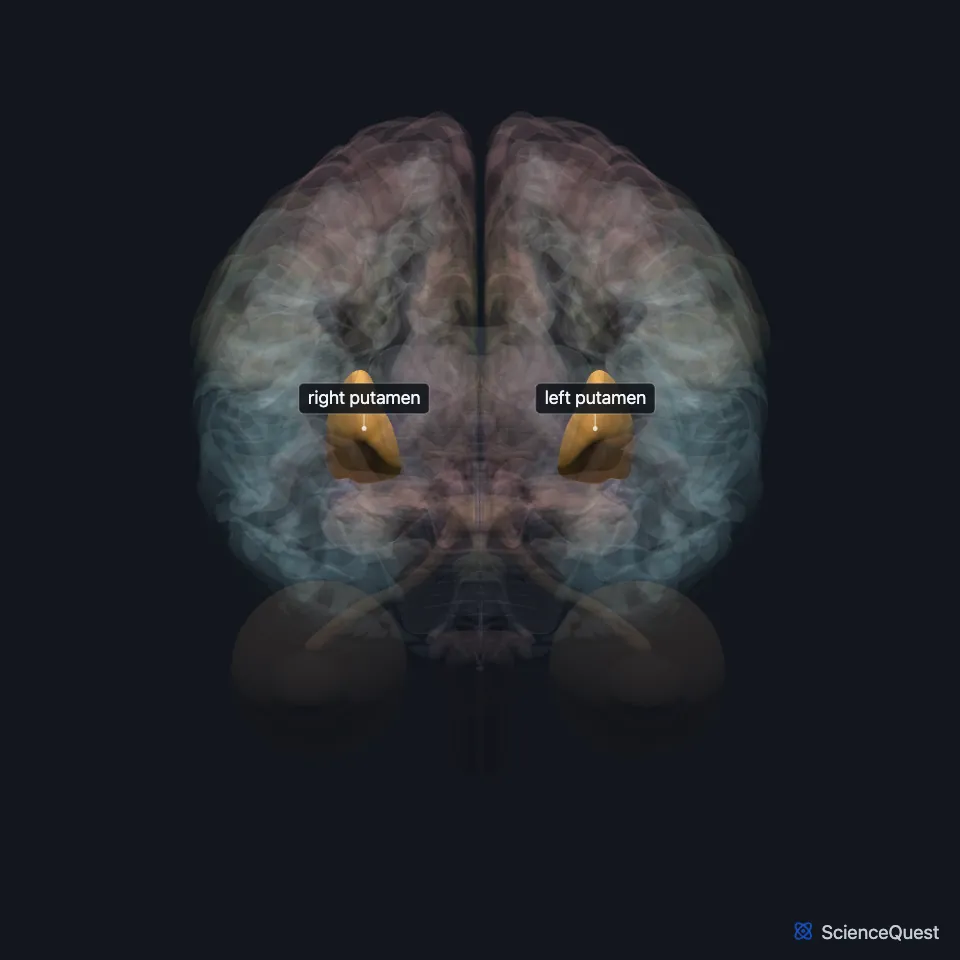
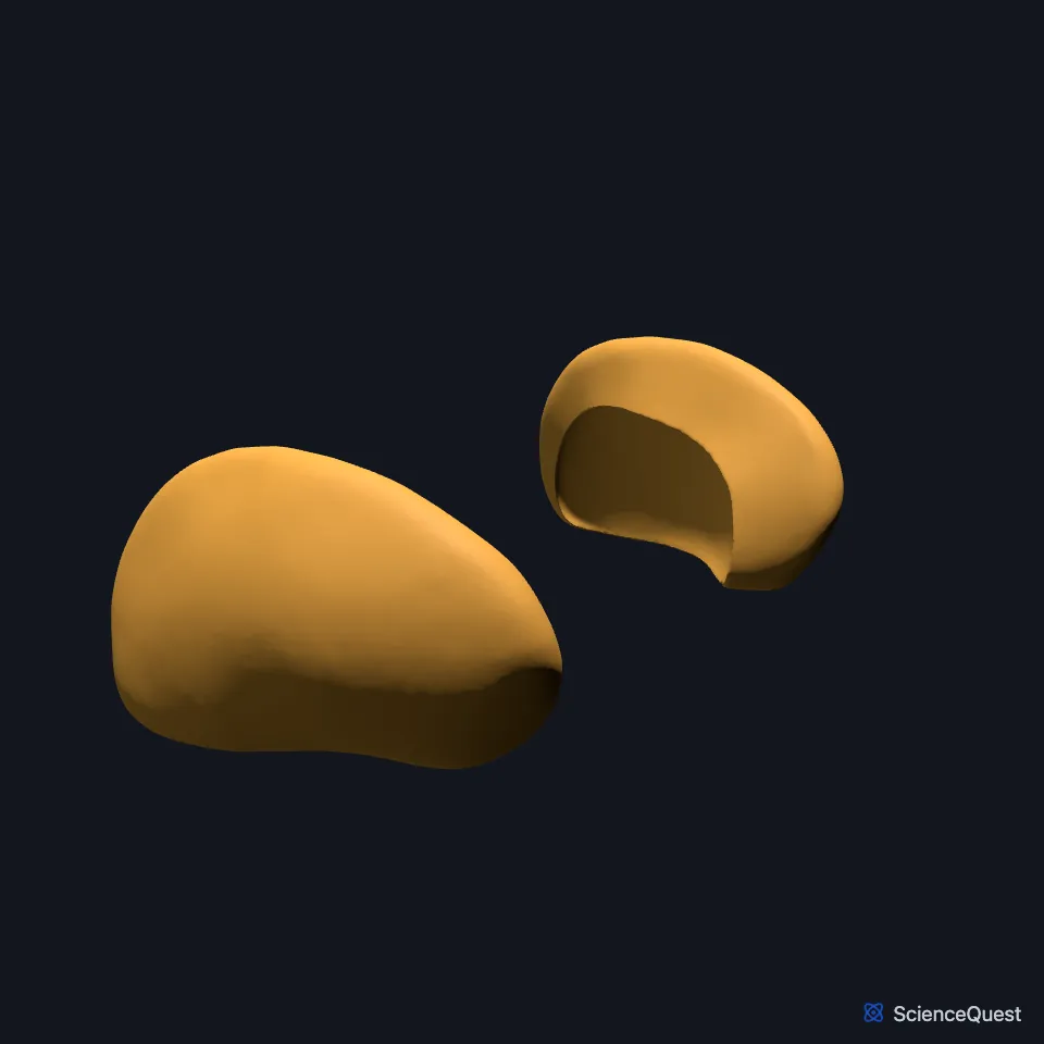
Where is the putamen?
It lies deep in the cerebral hemisphere, where it joins the globus pallidus to form the lentiform nucleus. (OpenStax A&P 2e, 13.2; StatPearls, Putamen)
In a frontal section it sits below the caudate nucleus, inside the curve of its head and body, with the internal capsule between them. The globus pallidus lies against its medial side. (OpenStax A&P 2e, 13.2; StatPearls, Globus Pallidus)
The lentiform nucleus that it forms with the globus pallidus lies beneath the insula. (StatPearls, Globus Pallidus)
What does the putamen do?
As part of the striatum it receives the cortex’s input to the basal nuclei, an excitatory connection that releases glutamate. (OpenStax A&P 2e, 13.2; StatPearls, Basal Ganglia)
From the striatum, the direct pathway runs to the internal segment of the globus pallidus and the substantia nigra pars reticulata. The indirect pathway runs through the external segment and the subthalamic nucleus to the same targets. (OpenStax A&P 2e, 13.2; StatPearls, Basal Ganglia)
Through the thalamus, the direct pathway ends up exciting the cortex and the indirect pathway ends up failing to excite it. (OpenStax A&P 2e, 13.2; StatPearls, Basal Ganglia)
It works in learning and motor control, speech articulation among them, and in language, reward, cognition and addiction. It also modulates the sensory and motor sides of pain. (StatPearls, Putamen)
How does the putamen connect to the cortex?
Its front part connects with the associative regions of the cortex. Its back part connects with the primary motor cortex and the supplementary motor area. (StatPearls, Putamen)
The striatum sends its output back to the cortex through the thalamus. (StatPearls, Putamen)
Which arteries supply the putamen, and how does it develop?
Perforating branches of the anterior and middle cerebral arteries, the lenticulostriate arteries, supply it; which artery predominates varies. (StatPearls, Putamen)
It develops from the telencephalon, which gives rise to most of the basal ganglia, the caudate nucleus among them. (StatPearls, Putamen)
Touches in this model
Measured on the BodyParts3D meshes: surfaces within 2 mm of the putamen, nearest first, 12 of 13.
- Anterior limb of left internal capsule in contact
- Anterior limb of right internal capsule in contact
- Left caudate nucleus in contact
- Left insula in contact
- Right caudate nucleus in contact
- Right insula in contact
- White matter structure of cerebral hemisphere in contact
- Left globus pallidus 0.5 mm away
- Right globus pallidus 0.5 mm away
- Anterior part of left superior temporal gyrus 1.2 mm away
- Anterior part of right superior temporal gyrus 1.2 mm away
- Left lateral ventricle 1.9 mm away
Sources
- OpenStax Anatomy and Physiology 2e, section 13.2, The Central Nervous System. CC BY-NC-SA 4.0. Read 2026-10-25.
- StatPearls, Neuroanatomy, Putamen. Mehrnoosh Ghandili; Sunil Munakomi. Last update January 30, 2023. CC BY-NC-ND 4.0. Read 2026-10-25.
- StatPearls, Neuroanatomy, Globus Pallidus. Nismat Javed; Marco Cascella. Last update February 20, 2023. CC BY-NC-ND 4.0. Read 2026-10-25.
- StatPearls, Neuroanatomy, Basal Ganglia. Carly B. Young; Vamsi Reddy; James Sonne. Last update July 24, 2023. CC BY-NC-ND 4.0. Read 2026-10-25.
The values are facts and free to use; this page’s selection and presentation are © 2026 ScienceQuest.