Precentral gyrus
The precentral gyrus is the gyrus of the frontal lobe that runs alongside the central sulcus. It contains the primary motor cortex. (Neuroscience Online, Overview of the Nervous System; StatPearls, Precentral Gyrus)
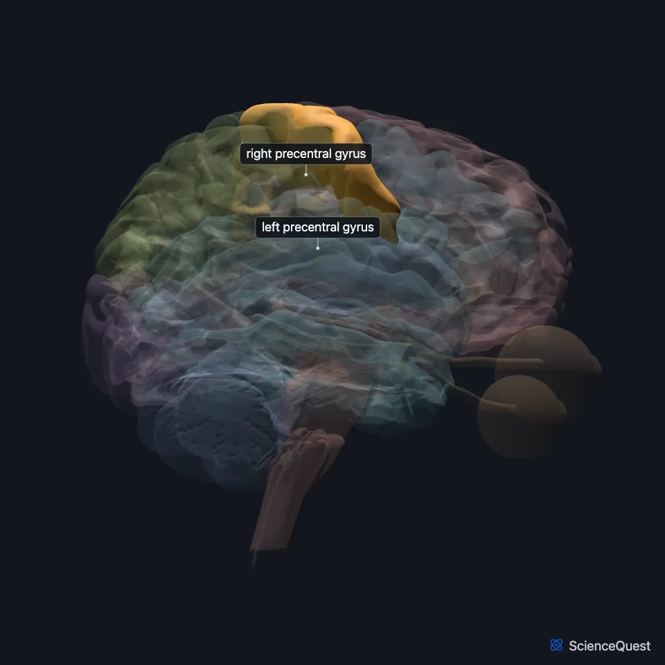
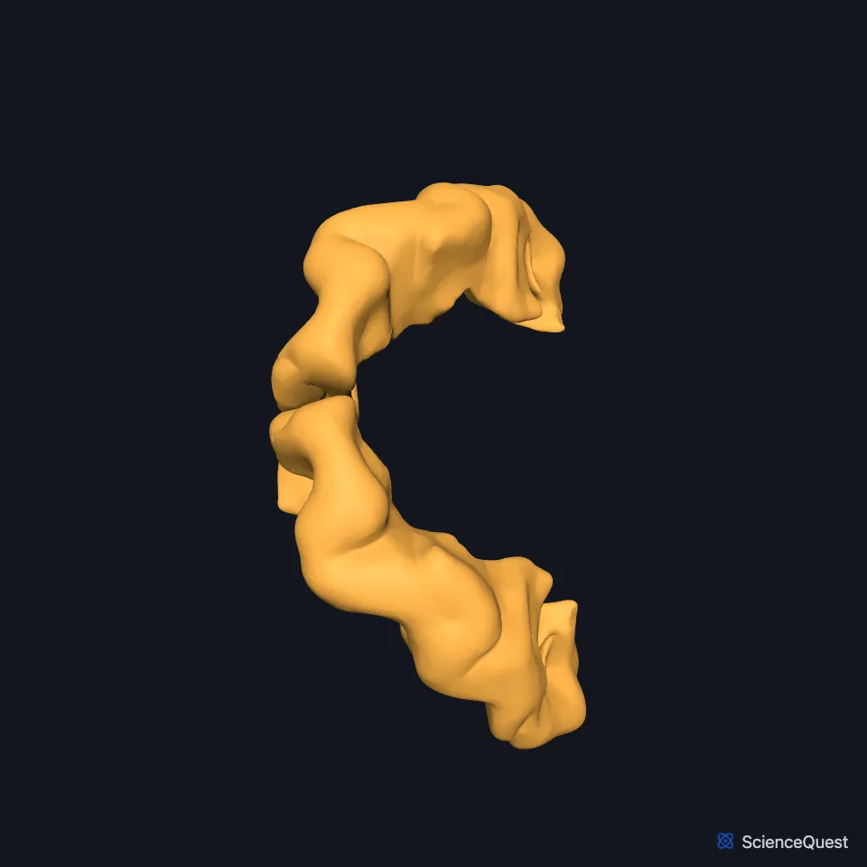
Where is the precentral gyrus?
It lies just in front of the central sulcus, the groove that separates the frontal lobe from the parietal lobe. The postcentral gyrus lies on the other side of that groove. (Neuroscience Online, Overview of the Nervous System and Motor Cortex; OpenStax Behavioral Neuroscience, 10.3)
The strip follows the central sulcus, which runs from the top edge of the hemisphere near its midpoint down and forward until it almost meets the lateral fissure, the groove above the temporal lobe. (OpenStax Behavioral Neuroscience, 10.3; Neuroscience Online, Overview of the Nervous System)
On the medial surface of the hemisphere, the primary motor cortex continues into the anterior paracentral lobule. (Neuroscience Online, Motor Cortex)
What does the precentral gyrus do?
Many of its output cells are upper motor neurons. Their axons run down to the brainstem or spinal cord, and in the spinal cord lower motor neurons carry the command on to the muscles. (OpenStax A&P 2e, 13.2; StatPearls, Motor Neuron and Precentral Gyrus)
It receives input from other cortical areas, the premotor cortex among them, and from the cerebellum and basal ganglia by way of the thalamus. Its corticospinal axons reach the alpha motor neurons of the spinal cord directly. (OpenStax A&P 2e, 14.3; Neuroscience Online, Motor Cortex)
Through the lateral corticospinal tract, each side’s motor cortex controls the arm and leg of the opposite side. Corticobulbar control of the head muscles is largely bilateral: most cranial motor nuclei get fibers from both sides, and only the lower face is served from the opposite side alone. The anterior corticospinal tract, which controls the trunk muscles, acts on both sides of the spinal cord. (OpenStax Behavioral Neuroscience, 10.3; OpenStax A&P 2e, 14.3; StatPearls, Corticobulbar Tract)
In Brodmann’s map the primary motor cortex is area 4. The premotor cortex lies just in front of it, in area 6 (Neuroscience Online adds area 8), and helps choose and prepare movements. (OpenStax A&P 2e, 16.2 and 13.2; Neuroscience Online, Motor Cortex and Higher Cortical Functions: Association and Executive Processing)
How is the body mapped on the precentral gyrus?
The gyrus carries a map of the body, the motor homunculus. The leg is represented on the medial surface, in the anterior paracentral lobule; moving down the lateral surface you pass the trunk, arm and hand, and the face sits farthest out. (OpenStax A&P 2e, 14.3; Neuroscience Online, Motor Cortex)
The hands and face, which make fine, exact movements, get far more cortex than the trunk or legs, whose movements are coarser. (OpenStax A&P 2e, 14.3; Neuroscience Online, Motor Cortex)
Unlike the primary sensory areas, this cortex has no cell-packed granular layer 4. Its most distinctive layer is the output layer 5, which holds the giant Betz cells. (Neuroscience Online, Motor Cortex)
Where do signals from the precentral gyrus go?
Among the tracts that begin here are the corticobulbar tract to the brainstem and the corticospinal tract to the spinal cord, whose fibers synapse on lower motor neurons. StatPearls also lists a cortico-rubrospinal tract. (OpenStax A&P 2e, 14.3; StatPearls, Precentral Gyrus, Lateral Corticospinal Tract and Motor Neuron)
Primary motor cortex neurons supply about 30% of the fibers in the corticospinal tract. The premotor and supplementary motor areas supply about 30%, the somatosensory cortex about 30% and the posterior parietal cortex about 10%. (Neuroscience Online, Motor Cortex)
The motor cortex also feeds the cerebellum: corticopontine fibers end in the pontine nuclei, which project to the cerebellum through its middle peduncle. With input from proprioceptors, the cerebellum adjusts motor commands for shifts in body position or load. (OpenStax A&P 2e, 14.3; Neuroscience Online, Motor Cortex and Cerebellum)
Where the sources differ
Sources describe which side these tracts serve differently. OpenStax A&P 2e section 14.3 calls corticobulbar axons ipsilateral, while OpenStax Behavioral Neuroscience 10.3 and StatPearls call the tract largely bilateral. For the anterior corticospinal tract, A&P 14.3 says its axons cross at the spinal level they serve and send some branches to the same side, and Behavioral Neuroscience 10.3 says most of it serves the same side. (OpenStax A&P 2e, 14.3; OpenStax Behavioral Neuroscience, 10.3; StatPearls, Corticobulbar Tract)
| Feature | Detail and source |
|---|---|
| Lobe | Frontal (Neuroscience Online, Overview of the Nervous System) |
| Cortex | Primary motor cortex (M1) (Neuroscience Online, Motor Cortex) |
| Brodmann area | 4 (OpenStax A&P 2e, 16.2; Neuroscience Online, Motor Cortex) |
| Output tracts | Corticospinal, corticobulbar and cortico-rubrospinal (OpenStax A&P 2e, 14.3; StatPearls, Precentral Gyrus) |
Touches in this model
Measured on the BodyParts3D meshes: surfaces within 2 mm of the precentral gyrus, nearest first, 12 of 22.
- Anterior part of left superior temporal gyrus in contact
- Anterior part of right superior temporal gyrus in contact
- Left accessory short gyrus in contact
- Left cingulate gyrus in contact
- Left insula in contact
- Left middle frontal gyrus in contact
- Left postcentral gyrus in contact
- Left superior frontal gyrus in contact
- Left superior parietal lobule and precuneus in contact
- Posterior part of left superior temporal gyrus in contact
- Posterior part of right superior temporal gyrus in contact
- Right accessory short gyrus in contact
Sources
- Neuroscience Online, McGovern Medical School, Section 2, Chapter 1, Overview of the Nervous System. Nachum Dafny, Ph.D.. Last update Reviewed and revised 07 Oct 2020. Open access; no reuse licence stated. Read 2026-10-25.
- StatPearls, Neuroanatomy, Precentral Gyrus. Linnea Banker; Prasanna Tadi. Last update July 24, 2023. CC BY-NC-ND 4.0. Read 2026-10-25.
- OpenStax Introduction to Behavioral Neuroscience, section 10.3, Our Brain Gets Involved: Responsibilities of Upper Motor Systems. CC BY-NC-SA 4.0. Read 2026-10-25.
- Neuroscience Online, McGovern Medical School, Section 3, Chapter 3, Motor Cortex. James Knierim, Ph.D.. Last update Last Review 07 July 2026. Open access; no reuse licence stated. Read 2026-10-25.
- OpenStax Anatomy and Physiology 2e, section 13.2, The Central Nervous System. CC BY-NC-SA 4.0. Read 2026-10-25.
- StatPearls, Neuroanatomy, Motor Neuron (introduction, read as the abstract on Europe PMC, PMID 32119503). Podraza LC, Hierholzer A, La Rose J. Abstract read on Europe PMC; no reuse licence stated. Read 2026-10-25.
- OpenStax Anatomy and Physiology 2e, section 14.3, Motor Responses. CC BY-NC-SA 4.0. Read 2026-10-25.
- StatPearls, Neuroanatomy, Corticobulbar Tract. Nikha Bhardwaj; Sisira Yadala. Last update July 24, 2023. CC BY-NC-ND 4.0. Read 2026-10-25.
- OpenStax Anatomy and Physiology 2e, section 16.2, The Mental Status Exam. CC BY-NC-SA 4.0. Read 2026-10-25.
- Neuroscience Online, McGovern Medical School, Section 4, Chapter 9, Higher Cortical Functions: Association and Executive Processing. Anthony Wright, Ph.D.. Last update Last Review 07 July 2026. Open access; no reuse licence stated. Read 2026-10-25.
- StatPearls, Neuroanatomy, Lateral Corticospinal Tract (introduction, read as the abstract on Europe PMC, PMID 30521239). Javed K, Reddy V, Lui F. Abstract read on Europe PMC; no reuse licence stated. Read 2026-10-25.
- Neuroscience Online, McGovern Medical School, Section 3, Chapter 5, Cerebellum. James Knierim, Ph.D.. Last update Last Review 15 July 2026. Open access; no reuse licence stated. Read 2026-10-25.
The values are facts and free to use; this page’s selection and presentation are © 2026 ScienceQuest.