Superior temporal gyrus
The superior temporal gyrus is the top one of the three oblique gyri of the temporal lobe, below the lateral fissure. (Neuroscience Online, Overview of the Nervous System)
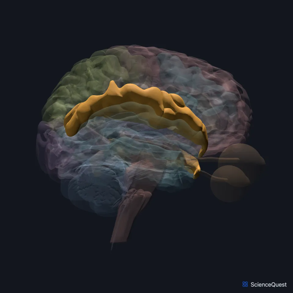
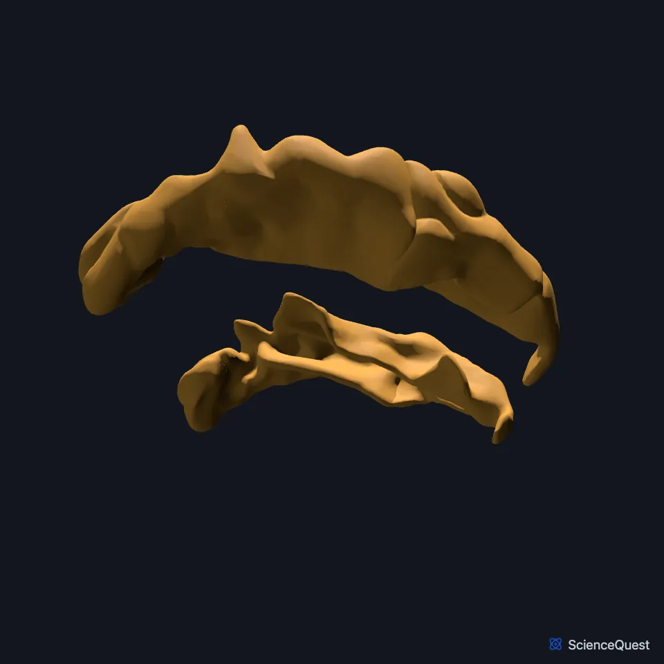
Where is the superior temporal gyrus?
Its upper surface extends into the lateral fissure, where it forms the transverse temporal gyrus of Heschl. (Neuroscience Online, Overview of the Nervous System)
Its caudal part reaches up to the parietal cortex. (Neuroscience Online, Overview of the Nervous System)
What does the superior temporal gyrus do?
It holds the primary auditory cortex, which OpenStax A&P numbers as Brodmann’s areas 41 and 42 in the superior temporal lobe. (Neuroscience Online, Overview of the Nervous System; OpenStax A&P 2e, 13.2)
Ascending auditory fibers stop in the inferior colliculus of the midbrain and the medial geniculate body of the thalamus before they reach the cortex of this gyrus. (Neuroscience Online, Auditory System: Pathways and Reflexes)
The frequency map of the ear holds all the way to the cortex. In primary auditory cortex, low frequencies lie more lateral. (Neuroscience Online, Auditory System: Pathways and Reflexes)
Its caudal part forms part of Wernicke’s area, which processes auditory information and matters for understanding language. (Neuroscience Online, Overview of the Nervous System; OpenStax Psychology 2e, 3.4; StatPearls, Wernicke Area)
What lies around the superior temporal gyrus, and what does it connect to?
Above the cochlear nuclei, auditory fibers run to both sides of the brain, so damage to one side’s pathway or auditory cortex does not usually cause deafness in either ear. (Neuroscience Online, Auditory System: Pathways and Reflexes; OpenStax A&P 2e, 14.2)
A deep white matter tract, the arcuate fasciculus, links Wernicke’s area with Broca’s area in the inferior frontal gyrus. Diffusion tractography also shows an indirect route that relays through the inferior parietal cortex. (OpenStax A&P 2e, 16.2; Catani et al. 2005, Ann Neurol; StatPearls, Wernicke Area)
The insula lies deep inside the lateral fissure, covered by the parts of the frontal, parietal and temporal lobes next to the sulcus, the operculum. (Neuroscience Online, Overview of the Nervous System)
Where the sources differ
OpenStax A&P 2e gives two answers for where primary auditory cortex sits in Brodmann’s map. Section 13.2 puts it in areas 41 and 42 in the superior temporal lobe; section 16.2 calls area 22 the primary auditory cortex. StatPearls instead assigns area 22 to Wernicke’s area. (OpenStax A&P 2e, 13.2 and 16.2; StatPearls, Wernicke Area)
Sources place Wernicke’s area differently. StatPearls puts it in Brodmann area 22, the posterior superior temporal gyrus of the dominant hemisphere, and OpenStax Psychology 2e in the temporal lobe. Neuroscience Online’s overview says the caudal superior temporal gyrus forms part of it, but it also calls the inferior parietal gyrus Wernicke’s area. (StatPearls, Wernicke Area; OpenStax Psychology 2e, 3.4; Neuroscience Online, Overview of the Nervous System)
| Feature | Detail and source |
|---|---|
| Lobe | Temporal (Neuroscience Online, Overview of the Nervous System) |
| Primary auditory cortex | Transverse temporal gyrus (of Heschl), on its upper surface (Neuroscience Online, Overview of the Nervous System) |
| Thalamic relay | Medial geniculate body (Neuroscience Online, Auditory System: Pathways and Reflexes) |
Touches in this model
Measured on the BodyParts3D meshes: surfaces within 2 mm of the superior temporal gyrus, nearest first, 12 of 27.
- Left accessory short gyrus in contact
- Left angular gyrus in contact
- Left inferior temporal gyrus in contact
- Left insula in contact
- Left middle temporal gyrus in contact
- Left postcentral gyrus in contact
- Left precentral gyrus in contact
- Left supramarginal gyrus in contact
- Orbital gyri and straight gyri, both sides in contact
- Right accessory short gyrus in contact
- Right angular gyrus in contact
- Right inferior temporal gyrus in contact
Sources
- Neuroscience Online, McGovern Medical School, Section 2, Chapter 1, Overview of the Nervous System. Nachum Dafny, Ph.D.. Last update Reviewed and revised 07 Oct 2020. Open access; no reuse licence stated. Read 2026-10-25.
- OpenStax Anatomy and Physiology 2e, section 13.2, The Central Nervous System. CC BY-NC-SA 4.0. Read 2026-10-25.
- Neuroscience Online, McGovern Medical School, Section 2, Chapter 13, Auditory System: Pathways and Reflexes. Lincoln Gray, Ph.D.. Last update Reviewed 20 July 2026. Open access; no reuse licence stated. Read 2026-10-25.
- OpenStax Psychology 2e, section 3.4, The Brain and Spinal Cord. CC BY-NC-SA 4.0. Read 2026-10-25.
- StatPearls, Neuroanatomy, Wernicke Area. Kinaan Javed; Vamsi Reddy; Joe M. Das; Michael Wroten. Last update July 24, 2023. CC BY-NC-ND 4.0. Read 2026-10-25.
- OpenStax Anatomy and Physiology 2e, section 14.2, Central Processing. CC BY-NC-SA 4.0. Read 2026-10-25.
- OpenStax Anatomy and Physiology 2e, section 16.2, The Mental Status Exam. CC BY-NC-SA 4.0. Read 2026-10-25.
- Annals of Neurology, Perisylvian language networks of the human brain, Ann Neurol 57, pages 8 to 16 (abstract, PMID 15597383). Catani M, Jones DK, ffytche DH. Abstract read on Europe PMC; no reuse licence stated. Read 2026-10-25.
The values are facts and free to use; this page’s selection and presentation are © 2026 ScienceQuest.