Fourth ventricle
The fourth ventricle is the lowest of the brain’s ventricles: the space between the cerebellum behind and the pons and upper medulla in front. (OpenStax A&P 2e, 13.3; StatPearls, Fourth Ventricle)
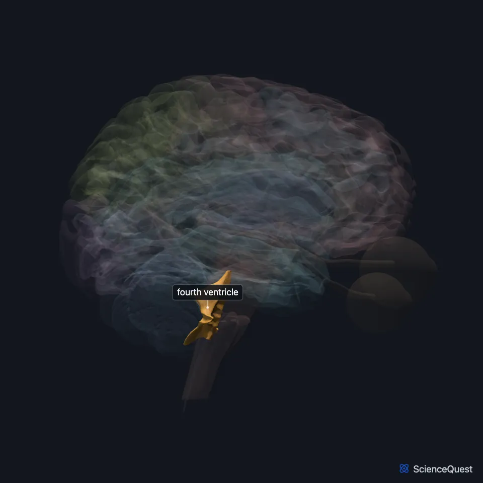
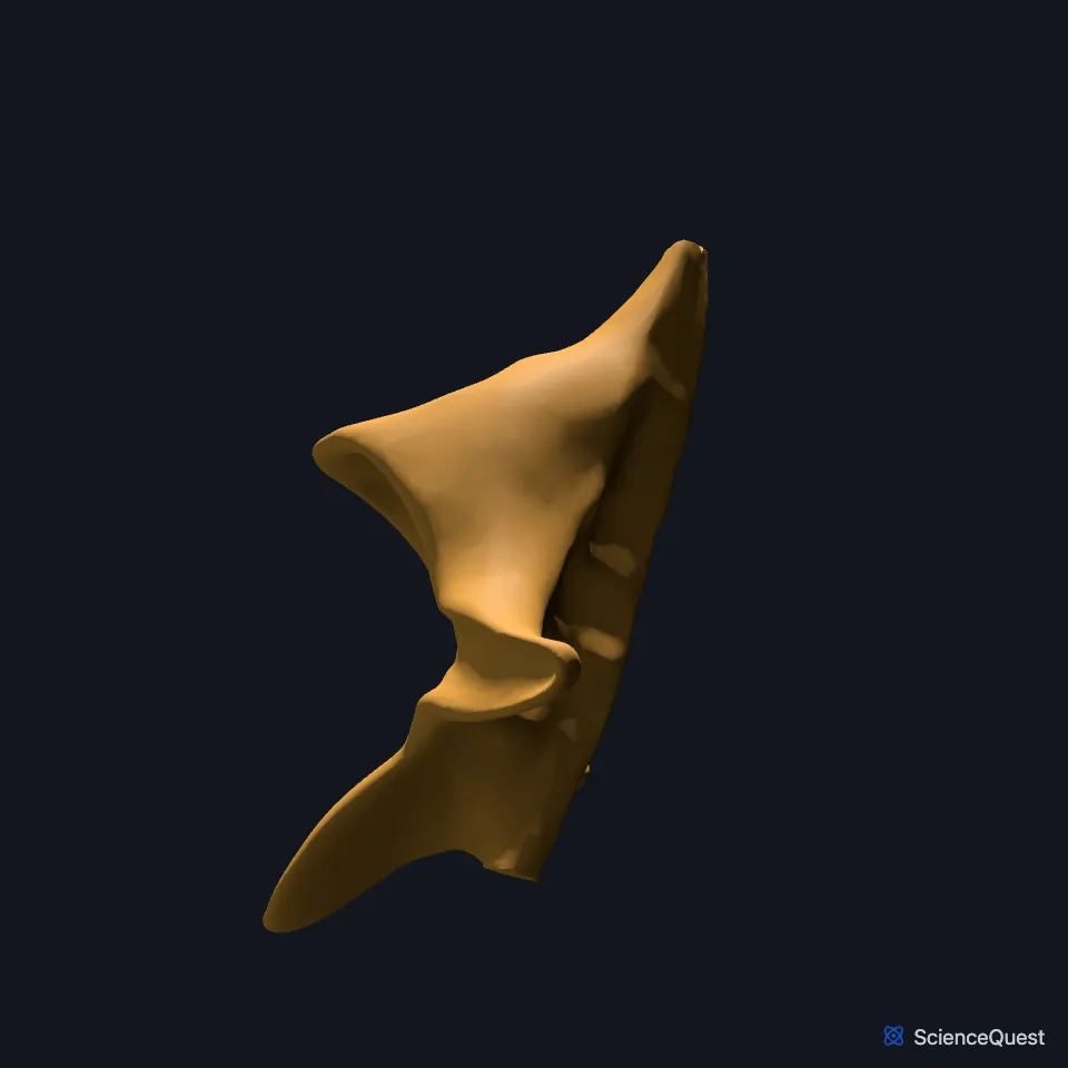
Where is the fourth ventricle?
The pons and medulla oblongata lie in front of it, the cerebellum behind it, and the spinal canal below it. (StatPearls, Fourth Ventricle)
Above, the cerebral aqueduct joins it to the third ventricle. Below, it narrows into the central canal of the spinal cord. (StatPearls, Fourth Ventricle; OpenStax A&P 2e, 13.3)
It forms from the cavity of the rhombencephalon, the hindbrain of the embryo. (StatPearls, Fourth Ventricle)
What forms the walls of the fourth ventricle?
Its floor is the back surface of the pons and upper medulla. (OpenStax A&P 2e, 13.3; StatPearls, Fourth Ventricle and Ventricular System)
Above, two cerebellar peduncles bridged by a thin sheet of white matter, the superior medullary velum, form its roof. The superior cerebellar peduncle bounds its side walls above; the inferior cerebellar peduncles and the cuneate and gracile tubercles bound them below. (StatPearls, Fourth Ventricle)
Lower down, the tela choroidea and the inferior medullary velum make up part of the roof. (StatPearls, Fourth Ventricle and Medulla Oblongata)
What does the fourth ventricle do?
Its own choroid plexus adds more CSF to the fluid that arrives through the aqueduct. (OpenStax A&P 2e, 13.3; StatPearls, Choroid Plexus, Fourth Ventricle and Cerebral Aqueduct (Sylvian))
Three openings let CSF out into the subarachnoid space around the brain and spinal cord: one median aperture, the foramen of Magendie, and two lateral apertures, the foramina of Luschka. (OpenStax A&P 2e, 13.3; StatPearls, Fourth Ventricle)
Fluid that does not leave through the apertures runs on down the central canal of the spinal cord. (OpenStax A&P 2e, 13.3; StatPearls, Fourth Ventricle)
What lies around the fourth ventricle?
The area postrema lies on its floor at the medulla, next to the solitary nucleus, where it detects emetic toxins in the blood and cerebrospinal fluid. It is also called the vomiting center, although a lesion of it does not stop vomiting from every emetic drug. (StatPearls, Medulla Oblongata)
It is the last ventricle in the chain, so a tumor or cyst blocking its apertures enlarges the whole ventricular system. The condition is called fourth ventricle outlet obstruction, a form of noncommunicating hydrocephalus. (StatPearls, Fourth Ventricle)
In one study cited by StatPearls, the posterior inferior, anterior inferior and superior cerebellar arteries supplied most of the blood to its choroid plexus. (StatPearls, Fourth Ventricle)
Touches in this model
Measured on the BodyParts3D meshes: surfaces within 2 mm of the fourth ventricle, nearest first.
- Central canal of spinal cord in contact
- Cerebellum in contact
- Cerebral aqueduct in contact
- Medulla oblongata in contact
- Midbrain in contact
- Pons in contact
- Left inferior colliculus 1.6 mm away
- Right inferior colliculus 1.6 mm away
Sources
- OpenStax Anatomy and Physiology 2e, section 13.3, Circulation and the Central Nervous System. CC BY-NC-SA 4.0. Read 2026-10-25.
- StatPearls, Neuroanatomy, Fourth Ventricle. Zachary K. Roesch; Prasanna Tadi. Last update August 7, 2023. CC BY-NC-ND 4.0. Read 2026-10-25.
- StatPearls, Neuroanatomy, Ventricular System. Saraswati Satyanarayan Shenoy; Forshing Lui. Last update July 24, 2023. CC BY-NC-ND 4.0. Read 2026-10-25.
- StatPearls, Neuroanatomy, Medulla Oblongata. Radostina Iordanova; Anil Kumar Reddy Reddivari. Last update July 24, 2023. CC BY-NC-ND 4.0. Read 2026-10-25.
- StatPearls, Neuroanatomy, Choroid Plexus. Kinaan Javed; Vamsi Reddy; Forshing Lui. Last update July 24, 2023. CC BY-NC-ND 4.0. Read 2026-10-25.
- StatPearls, Neuroanatomy, Cerebral Aqueduct (Sylvian). Jessica M. Rubino; Jeffery P. Hogg. Last update July 24, 2023. CC BY-NC-ND 4.0. Read 2026-10-25.
The values are facts and free to use; this page’s selection and presentation are © 2026 ScienceQuest.