Cerebellum
The cerebellum, Latin for “little brain”, lies behind the brain stem and the fourth ventricle, and its surface folds into many thin folia. (OpenStax A&P 2e, 13.2; StatPearls, Cerebellar Dysfunction)
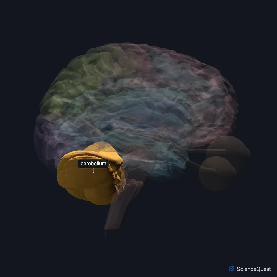
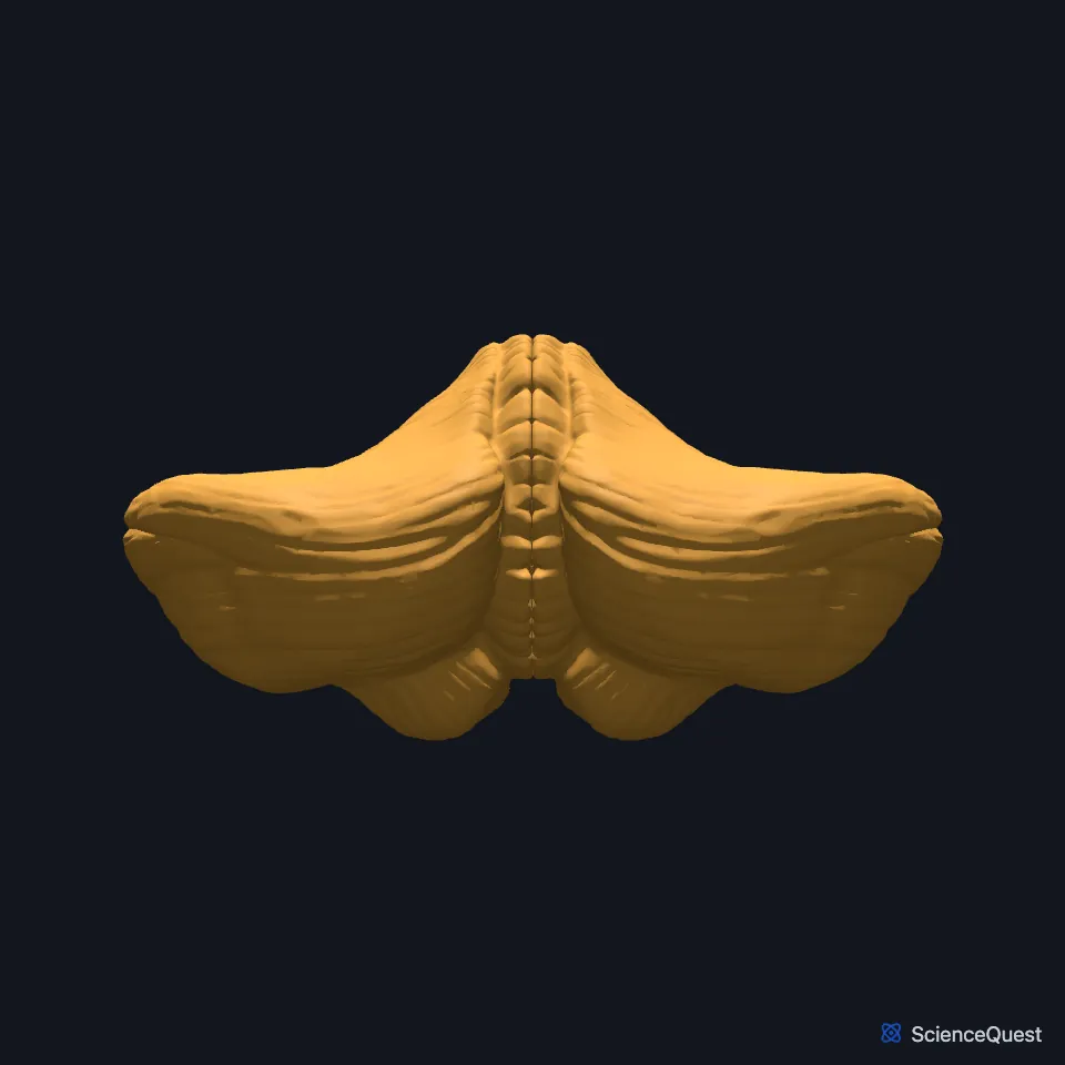
Where is the cerebellum?
It sits against the back of the brain stem, centered on the pons, in the posterior cranial fossa behind the fourth ventricle, the pons and the medulla oblongata. (OpenStax A&P 2e, 16.5; StatPearls, Cerebellum)
A tent-like shelf of dura, the tentorium cerebelli, separates it from the occipital lobes of the cerebrum above. (OpenStax A&P 2e, 13.3; StatPearls, Cerebellum)
What are the parts of the cerebellum?
Two hemispheres join at the midline vermis. The primary fissure separates the anterior and posterior lobes, and the posterolateral fissure separates the posterior and flocculonodular lobes. (StatPearls, Cerebellum)
A folded gray matter cortex covers a branched core of white matter, the arbor vitae, which surrounds three pairs of deep nuclei: from medial to lateral, the fastigial, interposed and dentate nuclei. (StatPearls, Cerebellum)
StatPearls puts 80% of the brain’s neurons in it, packed in a dense cell layer. (StatPearls, Cerebellum)
By function it divides into three zones. The spinocerebellum, the vermis and the paravermis beside it, takes body-sense input from the face and body; the vestibulocerebellum, the flocculonodular lobe, takes input from the vestibular nuclei; and the cerebrocerebellum, the rest of each hemisphere, takes input from the motor cortex through the pons. (OpenStax A&P 2e, 16.5; StatPearls, Cerebellar Dysfunction)
What does the cerebellum do?
It coordinates movement and keeps posture and balance. Its lateral hemispheres plan sequences of movement and take part in judging movement errors. (OpenStax A&P 2e, 13.2; StatPearls, Cerebellar Dysfunction and Cerebellum)
The midline regions keep balance and coordinate gait. The vermis coordinates the trunk, and the lateral hemispheres plan sequences of movement of the whole body. (OpenStax A&P 2e, 16.5; StatPearls, Cerebellum)
It also takes part in motor learning, how you learn and keep skilled movements. (OpenStax A&P 2e, 16.5; OpenStax Psychology 2e, 3.4; StatPearls, Cerebellar Dysfunction and Cerebellum)
Alcohol poisoning is the most common cause of cerebellar dysfunction. The resulting movement disorder, ataxia, shows as a wide, staggering gait and trouble walking heel to toe. (OpenStax A&P 2e, 14.3 and 16.5; StatPearls, Cerebellum and Cerebellar Dysfunction)
How does the cerebellum connect to the brain stem?
Three peduncles attach it: the superior to the midbrain, the middle to the pons, and the inferior to the medulla. (OpenStax A&P 2e, 16.5; StatPearls, Cerebellar Dysfunction, Cerebellum and Brainstem)
The largest of these links, the middle peduncle, joins it to the pons, and the pons and the cerebellum both develop from the metencephalon. (OpenStax A&P 2e, 13.1; StatPearls, Brainstem and Pons)
Where the sources differ
The sources disagree on whether the cerebellum starts movement. OpenStax A&P 2e says it is primarily responsible for initiating movements on the basis of equilibrium information; StatPearls says it cannot initiate muscle contraction. (OpenStax A&P 2e, 14.2; StatPearls, Cerebellum)
| Feature | Detail and source |
|---|---|
| Superior cerebellar peduncle | To the midbrain and forebrain; the main output, to the red nucleus and the thalamus (OpenStax A&P 2e, 16.5; StatPearls, Cerebellar Dysfunction) |
| Middle cerebellar peduncle | To the pons; carries the cortico-ponto-cerebellar pathway from the cerebral cortex, mainly to the lateral cerebellum (OpenStax A&P 2e, 16.5; StatPearls, Cerebellar Dysfunction) |
| Inferior cerebellar peduncle | To the medulla; its incoming fibers include the dorsal spinocerebellar, cuneocerebellar, olivocerebellar and vestibulocerebellar tracts (OpenStax A&P 2e, 16.5; StatPearls, Cerebellar Dysfunction) |
Touches in this model
Measured on the BodyParts3D meshes: surfaces within 2 mm of the cerebellum, nearest first, 12 of 17.
- Fourth ventricle in contact
- Left cingulate gyrus in contact
- Left inferior temporal gyrus in contact
- Pons in contact
- Right cingulate gyrus in contact
- Right inferior temporal gyrus in contact
- Left occipital lobe 0.3 mm away
- Right occipital lobe 0.3 mm away
- Left temporal bone 0.4 mm away
- Right temporal bone 0.5 mm away
- Medulla oblongata 0.7 mm away
- Occipital bone 0.8 mm away
Sources
- OpenStax Anatomy and Physiology 2e, section 13.2, The Central Nervous System. CC BY-NC-SA 4.0. Read 2026-10-25.
- StatPearls, Neuroanatomy, Cerebellar Dysfunction. Mahmut Unverdi; Khalid Alsayouri. Last update July 25, 2023. CC BY-NC-ND 4.0. Read 2026-10-25.
- OpenStax Anatomy and Physiology 2e, section 16.5, The Coordination and Gait Exams. CC BY-NC-SA 4.0. Read 2026-10-25.
- StatPearls, Neuroanatomy, Cerebellum. Sopiko Jimsheleishvili; Marine Dididze. Last update July 24, 2023. CC BY-NC-ND 4.0. Read 2026-10-25.
- OpenStax Anatomy and Physiology 2e, section 13.3, Circulation and the Central Nervous System. CC BY-NC-SA 4.0. Read 2026-10-25.
- OpenStax Psychology 2e, section 3.4, The Brain and Spinal Cord. CC BY-NC-SA 4.0. Read 2026-10-25.
- OpenStax Anatomy and Physiology 2e, section 14.3, Motor Responses. CC BY-NC-SA 4.0. Read 2026-10-25.
- StatPearls, Neuroanatomy, Brainstem. Hayden Basinger; Jeffery P. Hogg. Last update July 4, 2023. CC BY-NC-ND 4.0. Read 2026-10-25.
- OpenStax Anatomy and Physiology 2e, section 13.1, The Embryologic Perspective. CC BY-NC-SA 4.0. Read 2026-10-25.
- StatPearls, Neuroanatomy, Pons. Masum Rahman; Prasanna Tadi. Last update January 30, 2023. CC BY-NC-ND 4.0. Read 2026-10-25.
- OpenStax Anatomy and Physiology 2e, section 14.2, Central Processing. CC BY-NC-SA 4.0. Read 2026-10-25.
The values are facts and free to use; this page’s selection and presentation are © 2026 ScienceQuest.