Temporal bone
The temporal bones are a pair at the sides and base of the skull. Each holds the organs of hearing and balance, with the inner ear in its petrous part. (OpenStax A&P 2e, 7.2; Gray 1918, 5a. 4. The Temporal Bone; StatPearls, Temporal Region and Skull)
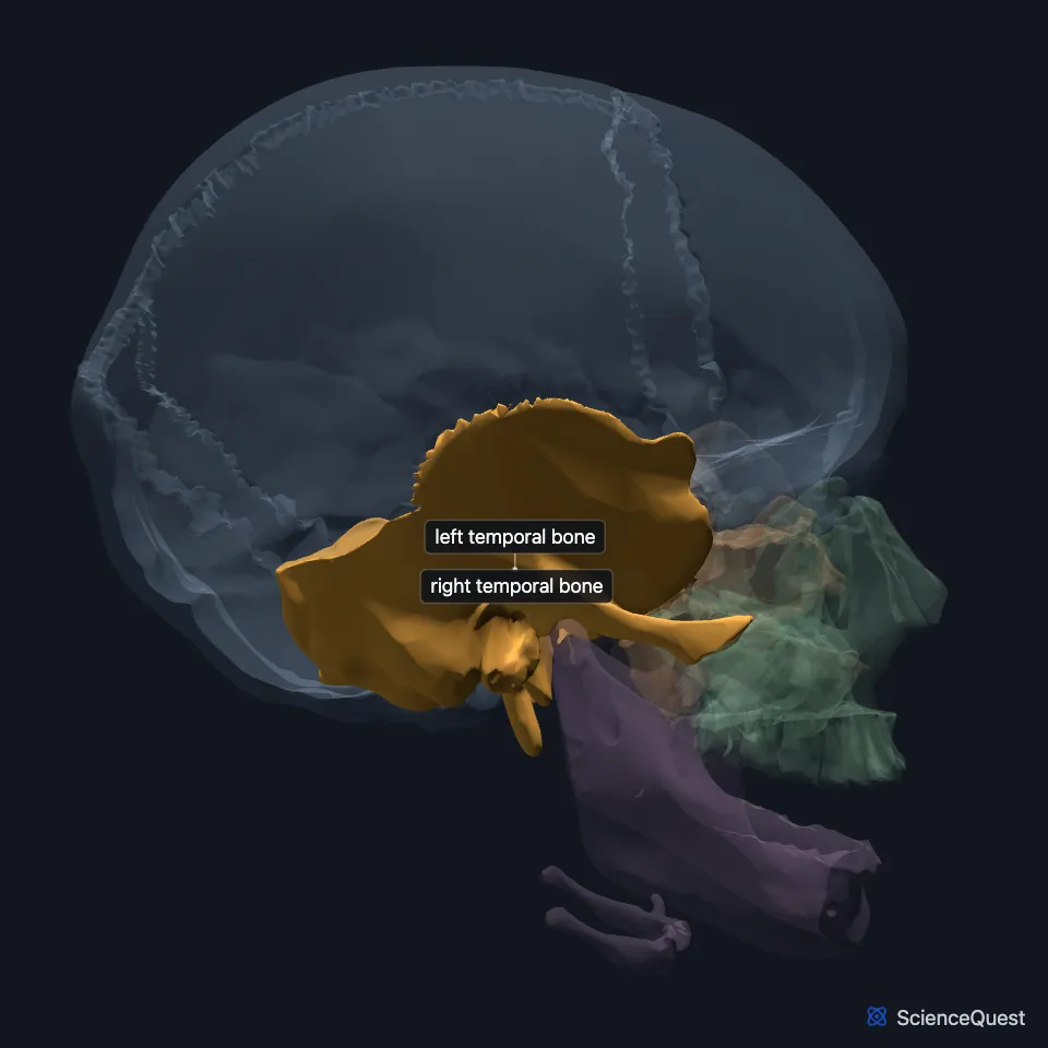
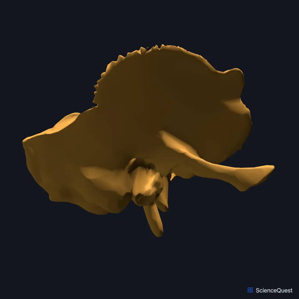
Where is the temporal bone?
Each temporal bone lies below a parietal bone, joined to it by the squamosal suture. Its mastoid part forms a bony prominence behind the auricle of the ear. (OpenStax A&P 2e, 7.2; StatPearls, Skull)
Inside the skull, the front surface of the petrous part forms the back of the middle cranial fossa. Its rear surface forms the front of the posterior cranial fossa. (OpenStax A&P 2e, 7.2; Gray 1918, 5a. 4. The Temporal Bone)
What are the parts of the temporal bone?
A long zygomatic process projects from the lower part of the squama, the thin upper part of the bone, and joins the zygomatic bone in the zygomatic arch. (OpenStax A&P 2e, 7.2; Gray 1918, 5a. 4. The Temporal Bone; StatPearls, Zygomatic)
Sound enters through the external auditory canal, the ear canal. Inside the skull, the internal acoustic meatus opens into the posterior cranial fossa and carries the facial nerve and the vestibulocochlear nerve, the nerve of hearing and balance. (OpenStax A&P 2e, 7.2; StatPearls, Temporal Region and Skull Foramen; Gray 1918, 5a. 4. The Temporal Bone)
The styloid process projects down from the underside of the petrous part. The stylohyoid, stylopharyngeus and styloglossus muscles attach to it, and so does the stylohyoid ligament, which runs to the lesser horn of the hyoid bone. (OpenStax A&P 2e, 7.2; StatPearls, Styloid Process)
What does the temporal bone do?
StatPearls names hearing and balance as its main functions. The ossicles and the cochlea serve hearing, and the three semicircular canals serve balance. (StatPearls, Temporal Region)
It carries the skull’s half of the jaw joint. The condyle of the mandible joins the mandibular fossa and the articular tubercle of the temporal bone, forming the temporomandibular joint that opens and closes the mouth. StatPearls places this fossa, which it calls the glenoid fossa, in the squamous part. (OpenStax A&P 2e, 7.2; StatPearls, Temporomandibular Joint)
Each internal carotid artery reaches the brain through a carotid canal in the temporal bone. Between the styloid and mastoid processes, the stylomastoid foramen ends the facial canal and lets the facial nerve out of the skull. (OpenStax A&P 2e, 7.2; StatPearls, Skull Foramen; Gray 1918, 5a. 4. The Temporal Bone)
What attaches to the temporal bone?
The mastoid process is a muscle attachment site. The sternocleidomastoid inserts on it. (OpenStax A&P 2e, 7.2; StatPearls, Foramen Magnum)
Its zygomatic process meets the temporal process of the zygomatic bone to form the zygomatic arch. The masseter arises from this arch. (OpenStax A&P 2e, 7.2; StatPearls, Mastication Muscles)
Behind, the mastoid part meets the occipital bone at the occipitomastoid suture. The jugular foramen lies at the junction of the occipital bone and the petrous part of the temporal bone. (StatPearls, Occipital Bone, Artery, Vein, and Nerve)
Above, the squamous suture unites its squamous part with the parietal bone. In front, it meets the sphenoid, whose greater wing shows on the side of the skull just ahead of the squamous part. (OpenStax A&P 2e, 7.2; StatPearls, Sphenoid Bone)
Where the sources differ
The sources divide the bone differently. StatPearls gives four parts, petrous, squamous, zygomatic and mastoid, and Gray’s Anatomy (1918) five: the squama, the petrous, mastoid and tympanic parts, and the styloid process. The Foundational Model of Anatomy lists squamous, petromastoid and tympanic parts and the styloid process, and OpenStax calls the zygomatic one a process. (StatPearls, Skull; Gray 1918, 5a. 4. The Temporal Bone; FMA, Temporal bone; OpenStax A&P 2e, 7.2)
| Feature | Detail and source |
|---|---|
| Parts | Squamous, petromastoid and tympanic parts, and the styloid process (FMA, Temporal bone) |
| Articulates with | Mandible (temporomandibular joint), parietal bone, occipital bone, zygomatic bone, sphenoid (OpenStax A&P 2e, 7.2; StatPearls, Occipital Bone, Artery, Vein, and Nerve and Sphenoid Bone) |
| Contains | The inner ear, in the petrous part (OpenStax A&P 2e, 7.2; StatPearls, Skull; Gray 1918, 5a. 4. The Temporal Bone) |
Touches in this model
Measured on the BodyParts3D meshes: surfaces within 2 mm of the temporal bone, nearest first, 12 of 37.
- Left parietal bone in contact
- Left zygomatic bone in contact
- Mandible in contact
- Occipital bone in contact
- Right parietal bone in contact
- Right zygomatic bone in contact
- Sphenoid bone in contact
- Deep part of left masseter in contact
- Deep part of right masseter in contact
- Left longissimus capitis in contact
- Left splenius capitis in contact
- Left sternocleidomastoid in contact
Sources
- OpenStax Anatomy and Physiology 2e, section 7.2, The Skull. CC BY-NC-SA 4.0. Read 2026-10-25.
- Gray’s Anatomy of the Human Body, 20th US edition (1918), II. Osteology, 5a. 4. The Temporal Bone. Henry Gray. Public domain. Read 2026-10-25.
- StatPearls, Anatomy, Head and Neck, Temporal Region. Arthur B. Dublin; Mohammed A. Al-Dhahir. Last update June 5, 2023. CC BY-NC-ND 4.0. Read 2026-10-25.
- StatPearls, Anatomy, Head and Neck, Skull. Bradley W. Anderson; Michael W. Kortz; Asa C. Black; Khalid A. Al Kharazi. Last update November 9, 2023. CC BY-NC-ND 4.0. Read 2026-10-25.
- StatPearls, Anatomy, Head and Neck, Zygomatic. Megan Yu; Shu-Min Wang. Last update June 5, 2023. CC BY-NC-ND 4.0. Read 2026-10-25.
- StatPearls, Anatomy, Head and Neck, Skull Foramen. Brody J. Lipsett; Khalid Alsayouri. Last update April 3, 2023. CC BY-NC-ND 4.0. Read 2026-10-25.
- StatPearls, Anatomy, Head and Neck, Styloid Process. Abdulaziz K. Abuhaimed; Reinier Alvarez; Ritesh G. Menezes. Last update January 9, 2023. CC BY-NC-ND 4.0. Read 2026-10-25.
- StatPearls, Anatomy, Head and Neck, Temporomandibular Joint. Bruno Bordoni; Melina Brizuela. Last update September 8, 2025. CC BY-NC-ND 4.0. Read 2026-10-25.
- StatPearls, Anatomy, Head and Neck: Foramen Magnum. Jennifer Ficke; Felix Jozsa. Last update April 11, 2026. CC BY-NC-ND 4.0. Read 2026-10-25.
- StatPearls, Anatomy, Head and Neck, Mastication Muscles. Hajira Basit; Muhammad Ali Tariq; Marco A. Siccardi. Last update June 5, 2023. CC BY-NC-ND 4.0. Read 2026-10-25.
- StatPearls, Anatomy, Head and Neck, Occipital Bone, Artery, Vein, and Nerve. Alex M. Germann; Varun Kashyap. Last update July 24, 2023. CC BY-NC-ND 4.0. Read 2026-10-25.
- StatPearls, Anatomy, Sphenoid Bone. Radia T. Jamil; Avery L. Callahan. Last update July 24, 2023. CC BY-NC-ND 4.0. Read 2026-10-25.
- Foundational Model of Anatomy 5.1.0, via EBI Ontology Lookup Service, Temporal bone (FMA52737), regional parts. FMA release terms, University of Washington. Read 2026-10-25.
The values are facts and free to use; this page’s selection and presentation are © 2026 ScienceQuest.