Hyoid bone
The hyoid is a small U-shaped bone in the midline of the neck, below the base of the mandible. It joins no other bone; muscles and tendons hold it in place. (OpenStax A&P 2e, 7.2; StatPearls, Hyoid Bone)
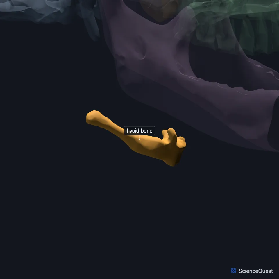
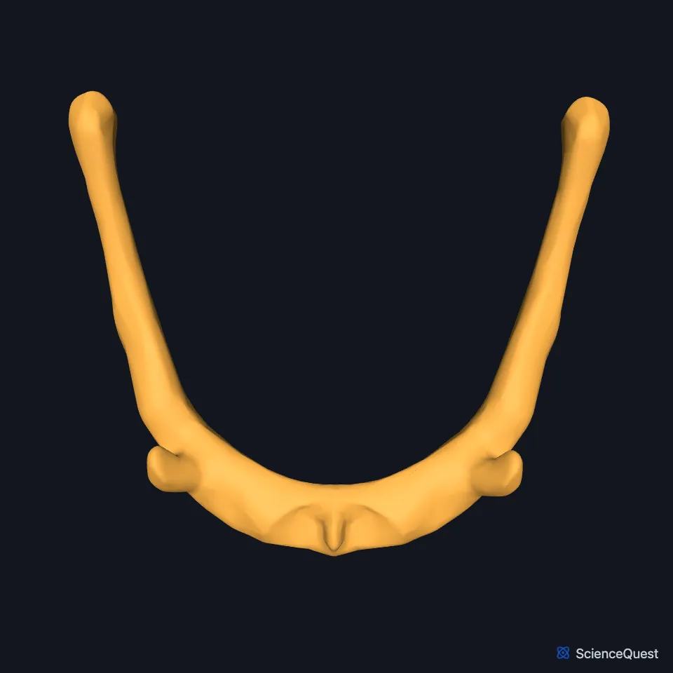
Where is the hyoid bone?
It lies in the midline of the neck, below the base of the mandible and in front of the fourth cervical vertebra. It sits just above the thyroid cartilage. In infancy it lies higher, in front of the second and third cervical vertebrae, and later in development it descends to the level of the fourth and fifth. (StatPearls, Hyoid Bone)
The tips of its U point backward. Muscles running to it from the larynx, pharynx, tongue and floor of the mouth anchor it in the anterior triangle of the neck. (OpenStax A&P 2e, 7.2; StatPearls, Hyoid Bone)
What are the parts of the hyoid?
It has a body, two greater horns and two lesser horns. The body and the greater horns make the U, with the greater horns as its limbs. (StatPearls, Hyoid Bone)
The horns join the body by fibrous tissue or by a true synovial joint. These joints often fuse with age. (StatPearls, Hyoid Bone)
What does the hyoid bone do?
Muscles from the larynx, pharynx, tongue and floor of the mouth anchor it. Through its link to the larynx it takes part in speech, and it also works in tongue movement, chewing, swallowing and breathing. (OpenStax A&P 2e, 7.2; StatPearls, Hyoid Bone)
Its position helps keep the airway open between the oropharynx and the trachea. Through its link to the larynx, it also takes part in voice production. (StatPearls, Hyoid Bone)
What holds the hyoid bone in place?
The stylohyoid ligaments hang it from the styloid processes of the temporal bones. Below, the thyrohyoid membrane ties it to the thyroid cartilage and limits how far it drops. (StatPearls, Hyoid Bone; OpenStax A&P 2e, 7.2)
Four pairs of suprahyoid muscles lie above it: the digastric, stylohyoid, mylohyoid and geniohyoid. Their main action is to raise the hyoid. (StatPearls, Hyoid Bone)
Four pairs of infrahyoid muscles lie below it: the sternohyoid, sternothyroid, omohyoid and thyrohyoid. Their main action is to lower the hyoid, and the sternohyoid runs to it from the back of the manubrium of the sternum. (StatPearls, Hyoid Bone)
How does the hyoid vary?
StatPearls ranks the hyoid among the most polymorphic parts of the body. The commonest variation is fusion of the joints between the horns and the body. (StatPearls, Hyoid Bone)
| Feature | Detail and source |
|---|---|
| Parts | Body, two greater horns, two lesser horns (StatPearls, Hyoid Bone) |
| Articulates with | No other bone (OpenStax A&P 2e, 7.2; StatPearls, Hyoid Bone) |
| Level | In front of the fourth cervical vertebra (the second and third in infancy) (StatPearls, Hyoid Bone) |
Touches in this model
Measured on the BodyParts3D meshes: surfaces within 2 mm of the hyoid bone, nearest first, 12 of 19.
- Left geniohyoid in contact
- Left longus capitis in contact
- Left mylohyoid in contact
- Left omohyoid in contact
- Left sternohyoid in contact
- Left stylohyoid in contact
- Left thyrohyoid in contact
- Right geniohyoid in contact
- Right longus capitis in contact
- Right mylohyoid in contact
- Right omohyoid in contact
- Right sternohyoid in contact
Sources
- OpenStax Anatomy and Physiology 2e, section 7.2, The Skull. CC BY-NC-SA 4.0. Read 2026-10-25.
- StatPearls, Anatomy, Head and Neck: Hyoid Bone. Dr. Rohin Garg; Adegbenro O. Fakoya; Ritesh G. Menezes. Last update May 3, 2025. CC BY-NC-ND 4.0. Read 2026-10-25.
The values are facts and free to use; this page’s selection and presentation are © 2026 ScienceQuest.