Mandible
The mandible is the lower jaw and the only movable bone of the skull. StatPearls calls it the largest bone of the skull. (OpenStax A&P 2e, 7.2; StatPearls, Mandible)
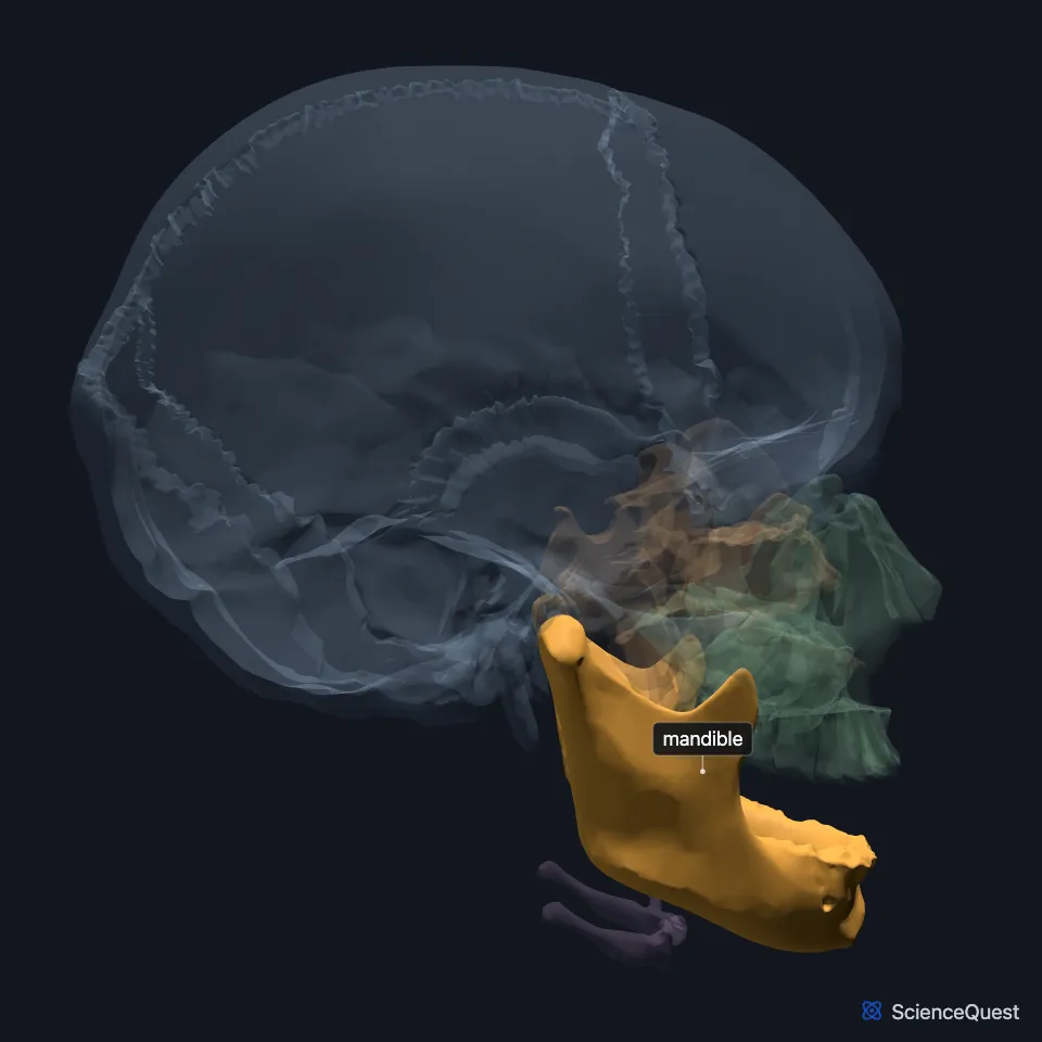
Where is the mandible?
It forms the lower jawline and the lower third of the face. It meets the skull base at the two temporomandibular joints, one on each side. (StatPearls, Mandible)
Each joint forms between the condyle of the mandible and the mandibular fossa of the temporal bone, with a fibrocartilage disc between them. (OpenStax A&P 2e, 7.2; StatPearls, Mandible)
What are the parts of the mandible?
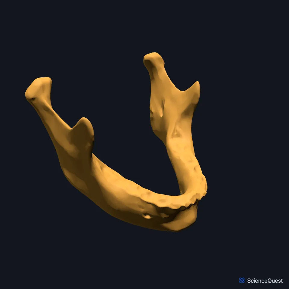
The mandible has a horizontal, U-shaped body and a vertical ramus on each side. They meet at the angle of the mandible. (OpenStax A&P 2e, 7.2; StatPearls, Mandible)
The upper border of each ramus ends in two processes, the coronoid in front and the condylar behind, which carries the condyle, or head, of the mandible. A deep curved notch, the mandibular notch, separates them. (OpenStax A&P 2e, 7.2; StatPearls, Mandible; Gray 1918, 5b. 8. The Mandible)
The upper surface of the body is alveolar bone lined with sockets for the lower teeth. At the front, below the midline symphysis, lies the mental protuberance, a triangular eminence at the chin. (OpenStax A&P 2e, 7.2; StatPearls, Mandible; Gray 1918, 5b. 8. The Mandible)
The mandibular foramen, on the inner side of the ramus, admits the inferior alveolar nerve and vessels to the mandibular canal. The canal runs through the body and ends at the mental foramen. (StatPearls, Mandible)
What does the mandible do?
The condyle joins the mandibular fossa and articular tubercle of the temporal bone to form the temporomandibular joint, which opens and closes the mouth. A biconcave fibrocartilage disc lies between the condyle and the fossa. (OpenStax A&P 2e, 7.2; StatPearls, Mandible)
In chewing, the mandible moves in five ways: elevation, depression, protrusion, retraction and side to side movement. (StatPearls, Mastication Muscles)
Which muscles attach to the mandible?
All four main muscles of chewing attach to the rami. The temporalis inserts on the coronoid process, the masseter on the outer surface of the ramus and angle, the medial pterygoid on the inner surface, and the lateral pterygoid on the neck of the condylar process. (StatPearls, Mastication Muscles and Mandible)
Muscles of the floor of the mouth and tongue arise from its inner surface: the mylohyoid from the mylohyoid line, the geniohyoid and genioglossus from the mental spine, and the anterior belly of the digastric from the digastric fossa. The mylohyoid and geniohyoid insert on the body of the hyoid bone, and the anterior belly of the digastric ends in an intermediate tendon attached to the hyoid. (StatPearls, Mandible and Hyoid Bone)
Facial muscles attach too. The platysma inserts on its lower border, the buccinator arises from the alveolar process, and the orbicularis oris arises from the incisive fossa. (StatPearls, Mandible)
How does the mandible change and vary?
At birth the mandible is a right and a left bone. They fuse during the first year at the mandibular symphysis into the single U-shaped adult bone. (OpenStax A&P 2e, 7.2; StatPearls, Mandible)
Male mandibles are generally squarer and more prominent than female ones. StatPearls puts this down to a larger mental protuberance and a smaller gonial angle in males. (StatPearls, Mandible)
Where the sources differ
The sources describe the mental protuberance differently. StatPearls calls it a midline depression enclosed where the ridge of the symphysis splits, while Gray’s Anatomy (1918) calls it a triangular eminence whose base is depressed in the center. (StatPearls, Mandible; Gray 1918, 5b. 8. The Mandible)
| Feature | Detail and source |
|---|---|
| Parts | Body, two rami, angles, coronoid and condylar processes (OpenStax A&P 2e, 7.2; StatPearls, Mandible) |
| Articulates with | Temporal bones, at the two temporomandibular joints (OpenStax A&P 2e, 7.2; StatPearls, Mandible) |
| Muscles of chewing | Temporalis, masseter, medial pterygoid, lateral pterygoid (StatPearls, Mastication Muscles) |
Touches in this model
Measured on the BodyParts3D meshes: surfaces within 2 mm of the mandible, nearest first, 12 of 52.
- Left temporal bone in contact
- Right temporal bone in contact
- Sphenoid bone in contact
- Anterior belly of left digastric in contact
- Anterior belly of right digastric in contact
- Deep part of left masseter in contact
- Deep part of right masseter in contact
- Gingiva of lower jaw in contact
- Left buccinator in contact
- Left depressor anguli oris in contact
- Left depressor labii inferioris in contact
- Left geniohyoid in contact
Sources
- OpenStax Anatomy and Physiology 2e, section 7.2, The Skull. CC BY-NC-SA 4.0. Read 2026-10-25.
- StatPearls, Anatomy, Head and Neck, Mandible. Grant Breeland; Aylin Aktar; Bhupendra C. Patel. Last update April 1, 2023. CC BY-NC-ND 4.0. Read 2026-10-25.
- Gray’s Anatomy of the Human Body, 20th US edition (1918), II. Osteology, 5b. 8. The Mandible (Lower Jaw) (1918 terms: condyloid process for condylar process). Henry Gray. Public domain. Read 2026-10-25.
- StatPearls, Anatomy, Head and Neck, Mastication Muscles. Hajira Basit; Muhammad Ali Tariq; Marco A. Siccardi. Last update June 5, 2023. CC BY-NC-ND 4.0. Read 2026-10-25.
- StatPearls, Anatomy, Head and Neck: Hyoid Bone. Dr. Rohin Garg; Adegbenro O. Fakoya; Ritesh G. Menezes. Last update May 3, 2025. CC BY-NC-ND 4.0. Read 2026-10-25.
The values are facts and free to use; this page’s selection and presentation are © 2026 ScienceQuest.