Parietal bone
The parietal bones are a right and left pair that form most of the upper side walls of the skull and meet at the top of the head. They are two of the eight bones of the brain case. (OpenStax A&P 2e, 7.2; Gray 1918, 5a. 2. The Parietal Bone and 5. The Skull)
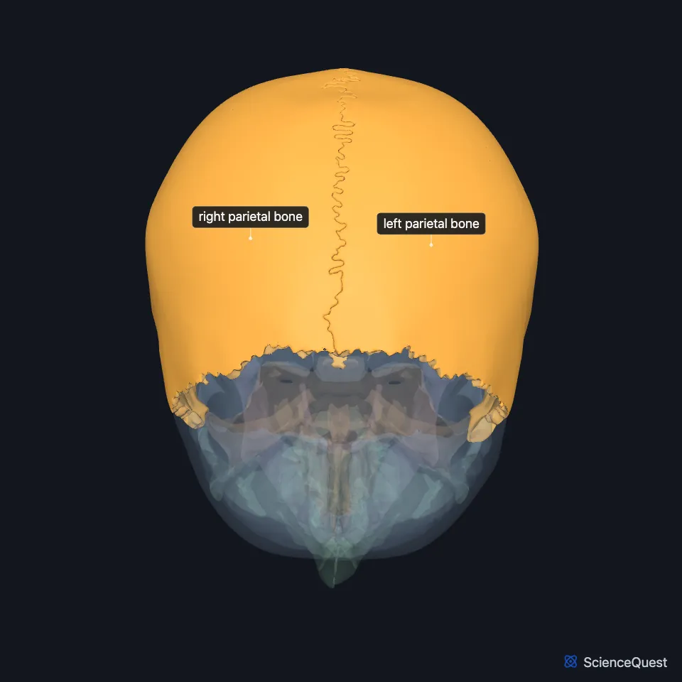
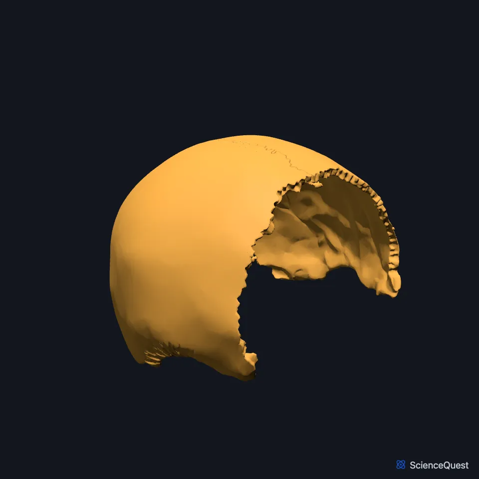
Where are the parietal bones?
Each parietal bone has the frontal bone in front, the temporal bone below and the occipital bone behind. (OpenStax A&P 2e, 7.2; StatPearls, Skull)
The parietal bones belong to the calvaria, the uppermost part of the skull, with the frontal, temporal and occipital bones. OpenStax groups the bones of the top and sides of the brain case as the flat bones of the skull. (StatPearls, Skull; OpenStax A&P 2e, 7.2)
What do the parietal bones do?
With the frontal, temporal and occipital bones, they form the calvaria, the top of the skull, which protects the brain. (OpenStax A&P 2e, 7.2; StatPearls, Skull)
Which sutures border the parietal bones?
The two parietal bones meet each other at the sagittal suture, which runs front to back along the top of the skull. In front, the coronal suture joins both to the frontal bone. (OpenStax A&P 2e, 7.2; StatPearls, Skull)
The squamous suture on the side of the skull joins each parietal bone to the temporal bone below. Behind, the lambdoid suture joins the parietal bones to the occipital bone; StatPearls names the occipital bone’s joint with the mastoid part of the temporal bone the occipitomastoid suture. (OpenStax A&P 2e, 7.2; StatPearls, Skull and Occipital Bone, Artery, Vein, and Nerve)
Each parietal bone takes part in two junctions on the side of the skull: the pterion, with the frontal, temporal and sphenoid bones, and the asterion, with the temporal and occipital bones. (StatPearls, Skull)
The front and back borders of each parietal bone are deeply serrated. Their teeth interlock with the frontal bone at the coronal suture and with the occipital bone at the lambdoid suture. (OpenStax A&P 2e, 7.2; Gray 1918, 5a. 2. The Parietal Bone)
How do the parietal bones change after birth?
In a newborn, the anterior fontanelle, the largest of the six fontanelles, lies where the two frontal bones meet the two parietal bones. The posterior fontanelle closes around 6 to 8 weeks of age and becomes the lambda, where the lambdoid and sagittal sutures meet. (StatPearls, Fontanelles and Skull)
Suture closure varies a great deal between people. Of the sutures that stay open into adulthood, StatPearls gives the sagittal as the first to close, around age 22, then the coronal, then the lambdoid around age 26. The metopic suture between the two halves of the frontal bone closes far earlier, usually at three months. (StatPearls, Skull)
Where the sources differ
Sources give different closing ages for the anterior fontanelle, between the frontal and parietal bones. StatPearls’ review of the fontanelles gives an average of 13 to 24 months; its review of the coronal suture gives 18 to 24 months. (StatPearls, Fontanelles and Coronal Suture)
| Feature | Detail and source |
|---|---|
| Paired | Yes, a right and a left parietal bone (OpenStax A&P 2e, 7.2; Gray 1918, 5. The Skull) |
| Articulates with | The other parietal bone, frontal bone, temporal bone, occipital bone, sphenoid (OpenStax A&P 2e, 7.2; StatPearls, Skull) |
| Sutures | Sagittal, coronal, squamous and lambdoid (StatPearls, Skull) |
Touches in this model
Measured on the BodyParts3D meshes: surfaces within 2 mm of the parietal bone, nearest first, 12 of 35.
- Frontal bone in contact
- Left temporal bone in contact
- Occipital bone in contact
- Right temporal bone in contact
- Sphenoid bone in contact
- Anterior part of left superior temporal gyrus in contact
- Anterior part of right superior temporal gyrus in contact
- Aponeurosis of epicranius in contact
- Left occipitalis in contact
- Left postcentral gyrus in contact
- Left superior frontal gyrus in contact
- Left superior parietal lobule and precuneus in contact
Sources
- OpenStax Anatomy and Physiology 2e, section 7.2, The Skull. CC BY-NC-SA 4.0. Read 2026-10-25.
- Gray’s Anatomy of the Human Body, 20th US edition (1918), II. Osteology, 5a. 2. The Parietal Bone. Henry Gray. Public domain. Read 2026-10-25.
- Gray’s Anatomy of the Human Body, 20th US edition (1918), II. Osteology, 5. The Skull, opening list of the cranial and facial bones. Henry Gray. Public domain. Read 2026-10-25.
- StatPearls, Anatomy, Head and Neck, Skull. Bradley W. Anderson; Michael W. Kortz; Asa C. Black; Khalid A. Al Kharazi. Last update November 9, 2023. CC BY-NC-ND 4.0. Read 2026-10-25.
- StatPearls, Anatomy, Head and Neck, Occipital Bone, Artery, Vein, and Nerve. Alex M. Germann; Varun Kashyap. Last update July 24, 2023. CC BY-NC-ND 4.0. Read 2026-10-25.
- StatPearls, Anatomy, Head and Neck: Fontanelles. Brody J. Lipsett; Vamsi Reddy; Kim Steanson. Last update July 24, 2023. CC BY-NC-ND 4.0. Read 2026-10-25.
- StatPearls, Anatomy, Head and Neck, Coronal Suture. William P. Russell; Mark R. Russell. Last update September 10, 2024. CC BY-NC-ND 4.0. Read 2026-10-25.
The values are facts and free to use; this page’s selection and presentation are © 2026 ScienceQuest.