Frontal bone
The frontal bone is the single bone of the forehead and the front and top of the skull. It is one of the eight bones of the brain case. (OpenStax A&P 2e, 7.2; StatPearls, Frontal Bone)
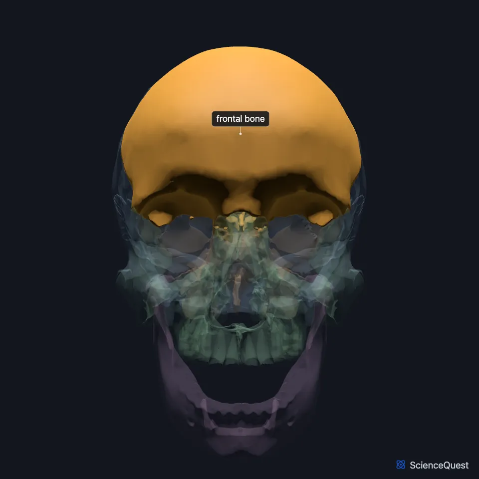
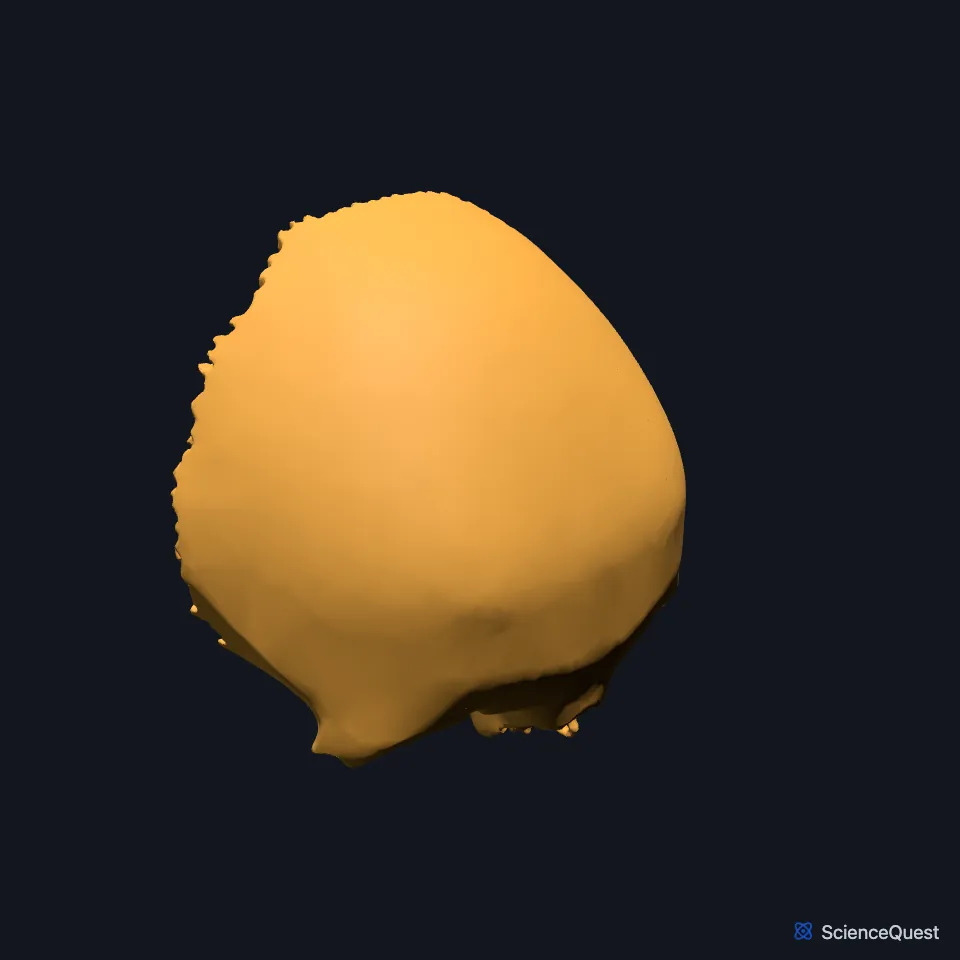
Where is the frontal bone?
Seen from the front, the frontal bone rises above the orbits as the forehead and forms the upper margin of each orbit, the supraorbital margin. (OpenStax A&P 2e, 7.2; StatPearls, Frontal Bone; Gray 1918, 5a. 3. The Frontal Bone)
Behind the supraorbital margins the bone turns back as two thin, triangular orbital plates. They roof the orbits, and their upper surface carries the frontal lobes of the brain in the anterior cranial fossa. (OpenStax A&P 2e, 7.2; StatPearls, Frontal Bone; Gray 1918, 5a. 3. The Frontal Bone)
The anterior cranial fossa holds the frontal lobes of the brain. The orbital plates of the frontal bone form its floor, together with the cribriform plate of the ethmoid and the small wings and front of the body of the sphenoid. (OpenStax A&P 2e, 7.2; StatPearls, Skull; Gray 1918, 5d. The Interior of the Skull)
What are the parts of the frontal bone?
The squamous part, the upper two thirds, is the forehead. Below it the nasal part meets the nasal bones at the nasion, and the orbital part runs back from the supraorbital margins as two triangular plates. (StatPearls, Frontal Bone)
Above each supraorbital margin the bone rises into a curved brow ridge, the superciliary arch, with a frontal sinus deep to it. The arches are generally larger in males than in females. (OpenStax A&P 2e, 7.2; Gray 1918, 5a. 3. The Frontal Bone; StatPearls, Skull)
Near the middle of each supraorbital margin, the supraorbital foramen passes a sensory nerve to the forehead. StatPearls describes supraorbital notches at the upper rim of the orbit that carry the supraorbital artery, veins and nerves. (OpenStax A&P 2e, 7.2; StatPearls, Frontal Bone)
Two frontal sinuses lie in the lower front part of the bone, above the orbital roofs. Their size varies widely from person to person. (OpenStax A&P 2e, 7.2; StatPearls, Frontal Bone)
What does the frontal bone do?
With the other bones of the brain case, the frontal bone encloses and protects the brain. It also forms the upper part of each orbit. (OpenStax A&P 2e, 7.2; StatPearls, Frontal Bone)
On its side, the frontal bone forms part of the front of the temporal fossa, the space the temporalis muscle occupies. Three muscles of facial expression act over the bone: the frontalis, the procerus and the orbicularis oculi. (StatPearls, Frontal Bone)
The ophthalmic branch of the trigeminal nerve supplies the skin over the bone. The facial nerve supplies the muscles over it. (StatPearls, Frontal Bone)
Which bones does the frontal bone meet?
The coronal suture joins the frontal bone to the right and left parietal bones. Where it crosses the sagittal suture lies the bregma, which in infancy is the open anterior fontanelle. (OpenStax A&P 2e, 7.2; StatPearls, Frontal Bone)
On the face it meets the nasal bones at the nasion and the frontal processes of the maxillae beside them. Around the orbit it meets the lacrimal bones, the sphenoid and the zygomatic bones. (StatPearls, Frontal Bone)
Inside the skull it meets the ethmoid below and in the midline, and the sphenoid behind and below. (StatPearls, Frontal Bone)
On the side of the skull, the frontal, parietal, temporal and sphenoid bones meet at the pterion, an H-shaped junction just above the ear. A blow here can tear the middle meningeal artery and cause an epidural hematoma. (OpenStax A&P 2e, 7.2; StatPearls, Frontal Bone and Skull)
How does the frontal bone develop?
Early in life the frontal bone is two halves, divided by the frontal suture, which fuses in normal development. StatPearls calls this the metopic suture and gives three months as its usual closure, with up to nine months possible. (StatPearls, Frontal Bone and Skull)
Where the sources differ
OpenStax describes the glabella as a slight depression in the midline between the eyebrows. StatPearls describes it as a midline prominence in the lower squamous part. (OpenStax A&P 2e, 7.2; StatPearls, Frontal Bone)
OpenStax says the frontal sinus may be divided at the midline into two spaces or fused into one. StatPearls describes two frontal sinuses that vary widely in size. (OpenStax A&P 2e, 7.2; StatPearls, Frontal Bone)
| Feature | Detail and source |
|---|---|
| Part of | The brain case, one of its eight bones (OpenStax A&P 2e, 7.2; Gray 1918, 5. The Skull) |
| Articulates with | Parietal bones, nasal bones, maxillae, lacrimal bones, sphenoid, zygomatic bones, ethmoid (StatPearls, Frontal Bone) |
| Openings | Paired supraorbital foramina and the single foramen cecum (StatPearls, Frontal Bone) |
| Sinus | Frontal sinus, above the orbital roofs (StatPearls, Frontal Bone) |
Touches in this model
Measured on the BodyParts3D meshes: surfaces within 2 mm of the frontal bone, nearest first, 12 of 37.
- Ethmoid in contact
- Left lacrimal bone in contact
- Left maxilla in contact
- Left nasal bone in contact
- Left parietal bone in contact
- Left zygomatic bone in contact
- Right lacrimal bone in contact
- Right maxilla in contact
- Right nasal bone in contact
- Right parietal bone in contact
- Right zygomatic bone in contact
- Sphenoid bone in contact
Sources
- OpenStax Anatomy and Physiology 2e, section 7.2, The Skull. CC BY-NC-SA 4.0. Read 2026-10-25.
- StatPearls, Anatomy, Head and Neck: Frontal Bone. Omesh Singh; Matthew A. Varacallo. Last update April 27, 2025. CC BY-NC-ND 4.0. Read 2026-10-25.
- Gray’s Anatomy of the Human Body, 20th US edition (1918), II. Osteology, 5a. 3. The Frontal Bone. Henry Gray. Public domain. Read 2026-10-25.
- StatPearls, Anatomy, Head and Neck, Skull. Bradley W. Anderson; Michael W. Kortz; Asa C. Black; Khalid A. Al Kharazi. Last update November 9, 2023. CC BY-NC-ND 4.0. Read 2026-10-25.
- Gray’s Anatomy of the Human Body, 20th US edition (1918), II. Osteology, 5d. The Interior of the Skull, Upper Surface of the Base of the Skull, and the table of craniometric points (1918 terms: small wings and great wing for lesser and greater wings). Henry Gray. Public domain. Read 2026-10-25.
- Gray’s Anatomy of the Human Body, 20th US edition (1918), II. Osteology, 5. The Skull, opening list of the cranial and facial bones. Henry Gray. Public domain. Read 2026-10-25.
The values are facts and free to use; this page’s selection and presentation are © 2026 ScienceQuest.