Orbicularis oculi
The orbicularis oculi is the ring of muscle around the eye, set around the upper and lower eyelids. Its main work is closing the lids, and it also helps tears drain. (StatPearls, Orbicularis Oculi Muscle; OpenStax A&P 2e, 11.3)
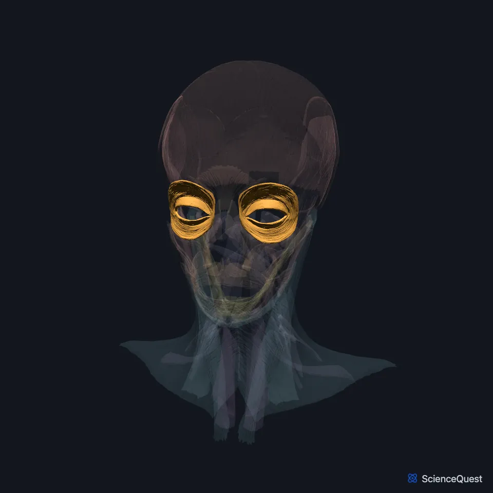
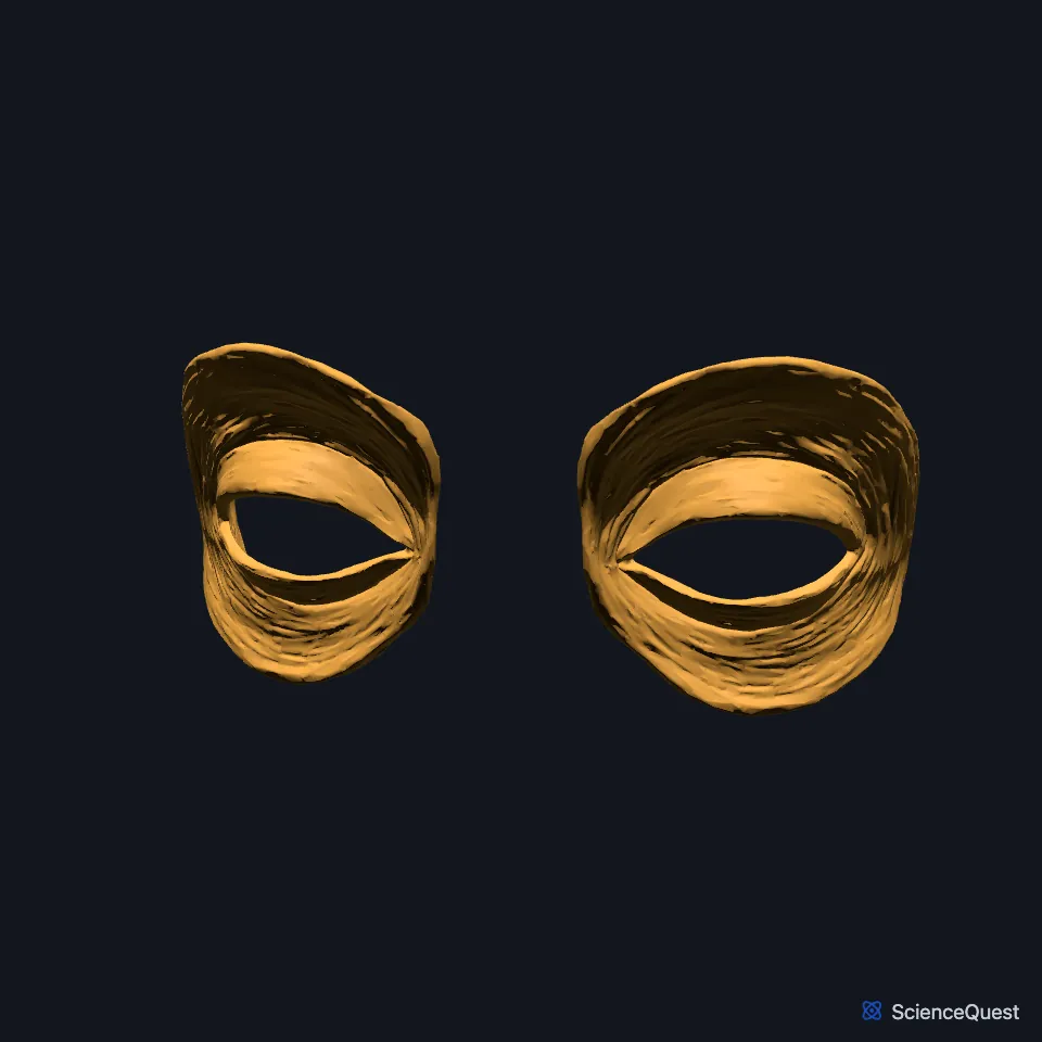
Open it in the Head and Neck Muscles Explorer See it alone in 3D
Where is the orbicularis oculi?
It lies just beneath the skin of the eyelids and stretches from the inner corner of the eye to the outer corner. (StatPearls, Orbicularis Oculi Muscle)
Almost no fat separates it from the skin. Its lower part contributes to the top of the cheek. (StatPearls, Facial Muscles and Cheeks)
What are the parts of the orbicularis oculi?
It has an orbital part and a palpebral part, each subdivided by function. (StatPearls, Orbicularis Oculi Muscle)
The orbital part attaches at the inner corner to the medial canthal tendon and the periosteum around it, and at the outer corner to the lateral palpebral raphe. At its edge it interweaves with neighboring muscles, among them the frontalis. (StatPearls, Orbicularis Oculi Muscle)
The palpebral part has preseptal, pretarsal and ciliary sections. The ciliary section, along the lid margin, shows as a colored line called the gray line. (StatPearls, Orbicularis Oculi Muscle)
About 88% of its fibers are fast-twitch, type II fibers. StatPearls names it and the zygomaticus major as the fastest-moving muscles in the body. (StatPearls, Orbicularis Oculi Muscle and Facial Muscles)
What does the orbicularis oculi do?
The orbital part closes the lids tight on purpose. The palpebral part does the blinking, both voluntary and involuntary. (StatPearls, Orbicularis Oculi Muscle)
Each blink squeezes the canaliculi, the small channels that carry tears to the lacrimal sac, and pulls the sac outward and forward. The negative pressure this creates draws tears into the sac and on down the nasolacrimal duct. (StatPearls, Orbicularis Oculi Muscle)
It also lowers the brows and makes the crow’s feet wrinkles at the outer corners of the eyes. (StatPearls, Facial Muscles)
Which nerve supplies the orbicularis oculi, and how does it form?
The facial nerve (cranial nerve VII) supplies it. Its temporal branch reaches the upper half, along with the frontalis and corrugator supercilii, and its zygomatic branch reaches the lower half. (StatPearls, Orbicularis Oculi Muscle and Facial Muscles)
When the muscle fails, as in Bell palsy, the eyelids no longer close as they should. (StatPearls, Orbicularis Oculi Muscle)
It develops from the mesenchyme of the second pharyngeal arch, the same arch as the orbicularis oris. (StatPearls, Orbicularis Oculi Muscle and Orbicularis Oris Muscle)
| Feature | Detail and source |
|---|---|
| Parts | Orbital and palpebral; the palpebral part has preseptal, pretarsal and ciliary sections (StatPearls, Orbicularis Oculi Muscle) |
| Origin | Medial bones of the orbit; medial canthal tendon (OpenStax A&P 2e, 11.3; StatPearls, Orbicularis Oculi Muscle) |
| Insertion | Lateral palpebral raphe, upper and lower tarsal plates, skin around the margin of the orbit (StatPearls, Orbicularis Oculi Muscle) |
| Action | Closes the eyelids; helps tears drain (StatPearls, Orbicularis Oculi Muscle) |
| Nerve | Facial nerve (CN VII): temporal and zygomatic branches (StatPearls, Orbicularis Oculi Muscle) |
Touches in this model
Measured on the BodyParts3D meshes: surfaces within 2 mm of the orbicularis oculi, nearest first, 12 of 26.
- Left corrugator supercilii in contact
- Left frontalis in contact
- Left levator labii superioris in contact
- Left levator labii superioris alaeque nasi in contact
- Left nasalis in contact
- Left zygomaticus minor in contact
- Right corrugator supercilii in contact
- Right frontalis in contact
- Right levator labii superioris in contact
- Right levator labii superioris alaeque nasi in contact
- Right nasalis in contact
- Right zygomaticus minor in contact
Sources
- StatPearls, Anatomy, Head and Neck: Orbicularis Oculi Muscle. John Tong; Michael J. Lopez; Adegbenro O. Fakoya; Bhupendra C. Patel. Last update May 25, 2024. CC BY-NC-ND 4.0. Read 2026-10-25.
- OpenStax Anatomy and Physiology 2e, section 11.3, Axial Muscles of the Head, Neck, and Back. CC BY-NC-SA 4.0. Read 2026-10-25.
- StatPearls, Anatomy, Head and Neck: Facial Muscles. Adegbenro O. Fakoya; Marc H. Hohman; Katherine E. Westbrook; Matthew A. Varacallo. Last update April 20, 2024. CC BY-NC-ND 4.0. Read 2026-10-25.
- StatPearls, Anatomy, Head and Neck, Cheeks. John D. Nguyen; Hieu Duong. Last update August 14, 2023. CC BY-NC-ND 4.0. Read 2026-10-25.
- StatPearls, Anatomy, Head and Neck, Orbicularis Oris Muscle. Prachi Jain; Manu Rathee. Last update August 7, 2023. CC BY-NC-ND 4.0. Read 2026-10-25.
The values are facts and free to use; this page’s selection and presentation are © 2026 ScienceQuest.