Zygomatic bone
The zygomatic bone is the cheekbone. Each of the pair forms the prominence of the cheek and part of the lateral wall and floor of the orbit. (OpenStax A&P 2e, 7.2; StatPearls, Zygomatic)
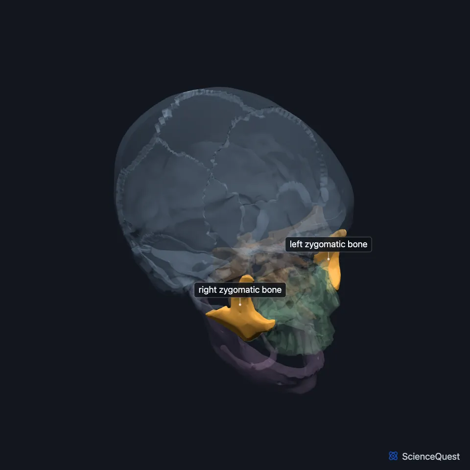
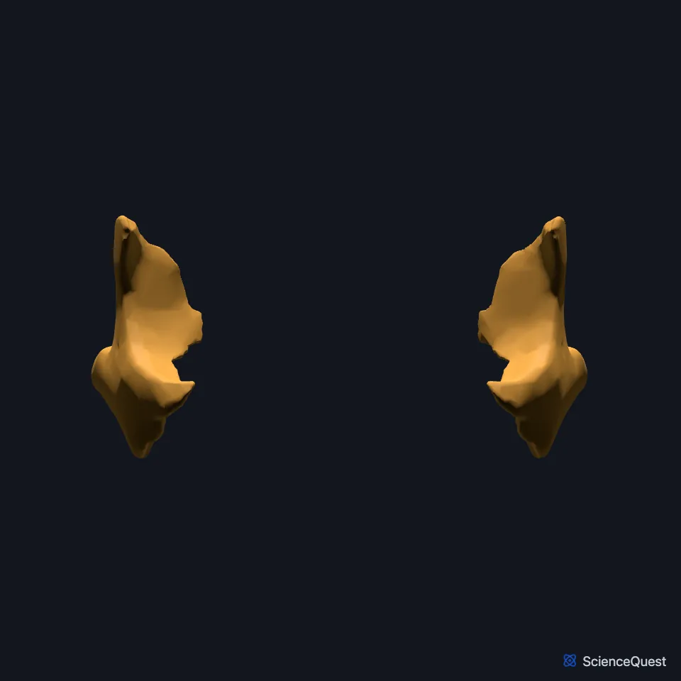
Where is the zygomatic bone?
The two bones project to the sides and form the prominence of the cheeks. Each also forms part of the orbital floor and parts of the temporal and infratemporal fossae. (StatPearls, Zygomatic)
Its temporal process runs back and joins the zygomatic process of the temporal bone to form the zygomatic arch. The masseter muscle arises from the arch. (OpenStax A&P 2e, 7.2; StatPearls, Zygomatic)
What does the zygomatic bone do?
It protects the contents of the orbit and shapes the contour of the face and cheek. (StatPearls, Zygomatic)
It withstands the forces of chewing and passes on forces from the maxilla. The zygomatic arch gives origin to the masseter, a chewing muscle that elevates the mandible. (StatPearls, Zygomatic and Mastication Muscles)
Which bones does the zygomatic bone meet?
It meets four bones: the frontal, the maxilla, the temporal and the sphenoid. (StatPearls, Zygomatic)
Its temporal process joins the zygomatic process of the temporal bone, and together they form the zygomatic arch. (OpenStax A&P 2e, 7.2; StatPearls, Zygomatic)
In the orbit it forms the lateral wall with the greater wing of the sphenoid, and part of the floor with the maxilla and palatine bone. (OpenStax A&P 2e, 7.2; StatPearls, Orbit Bones)
The zygomatic process of the maxilla articulates with the zygomatic bone and sets the malar eminence and the width of the face. (StatPearls, Maxilla)
How does the zygomatic bone vary?
Usually it is a single bone, but extra sutures can divide it into two or more parts. Most often one extra suture runs at right angles from the facial part of the maxillozygomatic suture, backward and outward across the bone, and splits it in two. (StatPearls, Zygomatic)
The zygomaticofacial foramen on its outer surface varies too. In a study of 102 dry half skulls it ranged from absent to four small openings, and half of the specimens had one. (StatPearls, Zygomatic)
| Feature | Detail and source |
|---|---|
| Articulates with | Frontal bone, maxilla, temporal bone, sphenoid (StatPearls, Zygomatic) |
| Forms | Cheek prominence, lateral orbital wall and part of its floor, front of the zygomatic arch (OpenStax A&P 2e, 7.2; StatPearls, Zygomatic) |
| Muscle origin | Masseter, from the zygomatic arch (StatPearls, Zygomatic) |
Touches in this model
Measured on the BodyParts3D meshes: surfaces within 2 mm of the zygomatic bone, nearest first, 12 of 24.
- Frontal bone in contact
- Left maxilla in contact
- Left temporal bone in contact
- Right maxilla in contact
- Right temporal bone in contact
- Sphenoid bone in contact
- Deep part of left masseter in contact
- Deep part of right masseter in contact
- Eyeball in contact
- Left levator labii superioris in contact
- Left zygomaticus major in contact
- Left zygomaticus minor in contact
Sources
- OpenStax Anatomy and Physiology 2e, section 7.2, The Skull. CC BY-NC-SA 4.0. Read 2026-10-25.
- StatPearls, Anatomy, Head and Neck, Zygomatic. Megan Yu; Shu-Min Wang. Last update June 5, 2023. CC BY-NC-ND 4.0. Read 2026-10-25.
- StatPearls, Anatomy, Head and Neck, Mastication Muscles. Hajira Basit; Muhammad Ali Tariq; Marco A. Siccardi. Last update June 5, 2023. CC BY-NC-ND 4.0. Read 2026-10-25.
- StatPearls, Anatomy, Head and Neck, Orbit Bones. Caleb L. Shumway; Mahsaw Motlagh; Matthew Wade. Last update July 24, 2023. CC BY-NC-ND 4.0. Read 2026-10-25.
- StatPearls, Anatomy, Head and Neck, Maxilla. Roberto M. Soriano; Melina Brizuela. Last update June 23, 2025. CC BY-NC-ND 4.0. Read 2026-10-25.
The values are facts and free to use; this page’s selection and presentation are © 2026 ScienceQuest.