Occipital lobe
The occipital lobe is the most caudal part of the brain. It contains the primary and higher-order visual cortex. (Neuroscience Online, Overview of the Nervous System)
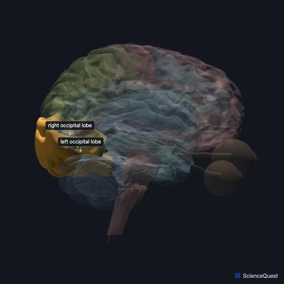
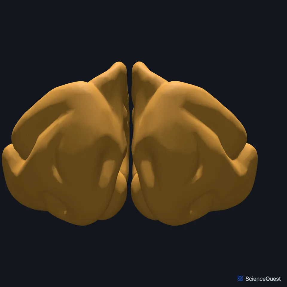
Where is the occipital lobe?
It rests on the tentorium cerebelli, which lies in the transverse fissure between the cerebral hemisphere and the cerebellum. (Neuroscience Online, Overview of the Nervous System)
On the medial surface, the parieto-occipital sulcus marks it off from the parietal lobe. The lateral surface has no such groove, so anatomists draw the border as an imaginary line from the tip of that sulcus to the preoccipital notch. (OpenStax A&P 2e, 13.2; Neuroscience Online, Overview of the Nervous System)
What are the parts of the occipital lobe?
On the medial surface, the calcarine fissure and the parieto-occipital sulcus define the cuneus, with the lingual gyrus below it. (Neuroscience Online, Overview of the Nervous System)
The primary visual cortex, Brodmann’s area 17, is the striate cortex. It straddles the calcarine fissure and wraps around the occipital pole onto the lateral side of the lobe. (Neuroscience Online, Visual Processing: Cortical Pathways; OpenStax A&P 2e, 16.2)
What does the occipital lobe do?
Areas 18 and 19, the extrastriate cortex around the primary visual cortex, carry out later stages of visual processing. (OpenStax A&P 2e, 16.2; Neuroscience Online, Visual Processing: Cortical Pathways)
In the primary visual cortex, most cells outside the color-sensitive blobs respond to either eye, and many fire best for an edge at one orientation. These cells start the analysis of shape. (OpenStax A&P 2e, 14.2; Neuroscience Online, Visual Processing: Cortical Pathways)
Each side’s visual cortex handles the opposite half of the visual field, and the map is upside down: the upper bank of the striate cortex receives the lower half of the field, and the lower bank the upper half. (OpenStax A&P 2e, 14.2; StatPearls, Visual Cortex; Neuroscience Online, Visual Processing: Cortical Pathways)
Where does visual information come from and go next?
The visual relay in the thalamus is the lateral geniculate nucleus, whose axons project to the visual cortex in the occipital lobe. (Neuroscience Online, Visual Processing: Cortical Pathways; OpenStax A&P 2e, 14.2)
The extrastriate cortex surrounds V1 in the occipital lobe, and its two main areas next to V1 are V2 and V3. StatPearls divides the visual cortex into five areas, V1 to V5, and V1 sends its color, shape, location and motion signals to separate parts of the extrastriate cortex. (OpenStax A&P 2e, 14.2; Neuroscience Online, Visual Processing: Cortical Pathways; StatPearls, Visual Cortex)
From there, processing splits in two. A ventral stream to the inferior temporal cortex identifies objects and colors, and a dorsal stream to the parietal cortex tracks where things are and guides your movements toward them. (OpenStax A&P 2e, 14.2; Neuroscience Online, Visual Processing: Cortical Pathways; Goodale and Milner 1992, Trends Neurosci)
Where the sources differ
Sources differ on which Brodmann areas make up the primary visual cortex. OpenStax A&P 2e section 13.2 says areas 17 and 18 are responsible for primary visual perception. Section 16.2 and Neuroscience Online give area 17 alone, with areas 18 and 19 as later stages. (OpenStax A&P 2e, 13.2 and 16.2; Neuroscience Online, Visual Processing: Cortical Pathways)
| Feature | Detail and source |
|---|---|
| Primary visual cortex | Brodmann’s area 17 (V1), the striate cortex (OpenStax A&P 2e, 16.2; Neuroscience Online, Visual Processing: Cortical Pathways) |
| Thalamic relay | Lateral geniculate nucleus (Neuroscience Online, Visual Processing: Cortical Pathways) |
| Medial landmarks | Calcarine fissure and parieto-occipital sulcus (Neuroscience Online, Overview of the Nervous System) |
Touches in this model
Measured on the BodyParts3D meshes: surfaces within 2 mm of the occipital lobe, nearest first, 12 of 21.
- Left angular gyrus in contact
- Left cingulate gyrus in contact
- Left fusiform gyrus in contact
- Left inferior temporal gyrus in contact
- Left lateral ventricle in contact
- Left middle temporal gyrus in contact
- Left superior parietal lobule and precuneus in contact
- Right angular gyrus in contact
- Right cingulate gyrus in contact
- Right fusiform gyrus in contact
- Right inferior temporal gyrus in contact
- Right lateral ventricle in contact
Sources
- Neuroscience Online, McGovern Medical School, Section 2, Chapter 1, Overview of the Nervous System. Nachum Dafny, Ph.D.. Last update Reviewed and revised 07 Oct 2020. Open access; no reuse licence stated. Read 2026-10-25.
- OpenStax Anatomy and Physiology 2e, section 13.2, The Central Nervous System. CC BY-NC-SA 4.0. Read 2026-10-25.
- Neuroscience Online, McGovern Medical School, Section 2, Chapter 15, Visual Processing: Cortical Pathways. Valentin Dragoi, Ph.D. (content provided by Chieyeko Tsuchitani, Ph.D.). Last update Reviewed 07 July 2026. Open access; no reuse licence stated. Read 2026-10-25.
- OpenStax Anatomy and Physiology 2e, section 16.2, The Mental Status Exam. CC BY-NC-SA 4.0. Read 2026-10-25.
- OpenStax Anatomy and Physiology 2e, section 14.2, Central Processing. CC BY-NC-SA 4.0. Read 2026-10-25.
- StatPearls, Neuroanatomy, Visual Cortex (introduction, read as the abstract on Europe PMC, PMID 29494110). Huff T, Mahabadi N, Tadi P. Abstract read on Europe PMC; no reuse licence stated. Read 2026-10-25.
- Trends in Neurosciences, Separate visual pathways for perception and action, Trends Neurosci 15, pages 20 to 25 (abstract, PMID 1374953). Goodale MA, Milner AD. Abstract read on Europe PMC; no reuse licence stated. Read 2026-10-25.
The values are facts and free to use; this page’s selection and presentation are © 2026 ScienceQuest.