Palatine bone
The palatine bone is a paired, L-shaped facial bone at the back of the nasal cavity, between the maxilla and the pterygoid process of the sphenoid. (Gray 1918, 5b. 5. The Palatine Bone; StatPearls, Skull)
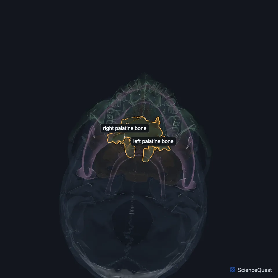
Where is the palatine bone?
Each one walls three cavities: the floor and side wall of the nasal cavity, the roof of the mouth and the floor of the orbit. It also forms part of the pterygopalatine and pterygoid fossae and of the inferior orbital fissure. (Gray 1918, 5b. 5. The Palatine Bone; StatPearls, Orbit Bones)
What are the parts of the palatine bone?
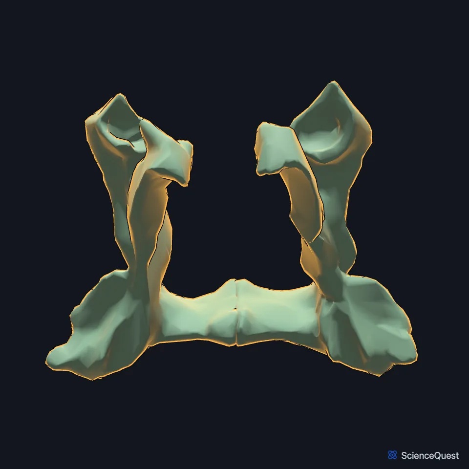
It has a horizontal part and a vertical part, joined like the two strokes of an L. Three processes stand out: the pyramidal process, pointing back and out from the corner, and the orbital and sphenoidal processes on top of the vertical part. (Gray 1918, 5b. 5. The Palatine Bone)
The notch between the orbital and sphenoidal processes is closed by the sphenoid into the sphenopalatine foramen. It leads from the pterygopalatine fossa into the nose and carries the sphenopalatine vessels and the superior nasal and nasopalatine nerves. (Gray 1918, 5b. 5. The Palatine Bone)
What does the palatine bone do?
Its horizontal plates close the back of the hard palate, which separates the mouth from the nose. In front of them, the palatine processes of the maxillae make up most of it. (StatPearls, Palate)
The free back edge of the horizontal part anchors the soft palate. Where the two bones meet there, they form the posterior nasal spine, which the muscle of the uvula attaches to. (Gray 1918, 5b. 5. The Palatine Bone)
It also passes nerves to the palate. The greater palatine vessels and nerve leave through the greater palatine foramen beside the third molar, and the lesser palatine nerve through the lesser palatine foramen on the pyramidal process. (StatPearls, Palate; Gray 1918, 5b. 5. The Palatine Bone)
Which bones meet the palatine bone?
Gray’s Anatomy (1918) lists six: the sphenoid, the ethmoid, the maxilla, the inferior nasal concha, the vomer and the palatine bone of the other side. (Gray 1918, 5b. 5. The Palatine Bone)
It bounds the pterygopalatine fossa, a space behind the maxilla. Through 8 foramina and canals the fossa connects with the orbit, nasal cavity, mouth, infratemporal fossa, middle cranial fossa and pharynx. StatPearls places the palatine bone in its floor and front inner wall. (StatPearls, Pterygopalatine Fossa)
How does the palatine bone grow?
Gray’s Anatomy (1918) describes it ossifying in membrane from one center at about the sixth to eighth week of fetal life, but notes that some authorities describe four. At birth its vertical part is about as tall as the horizontal part is wide; in the adult it is about twice as tall. (Gray 1918, 5b. 5. The Palatine Bone)
| Feature | Detail and source |
|---|---|
| Parts | Horizontal and vertical parts; pyramidal, orbital and sphenoidal processes (Gray 1918, 5b. 5. The Palatine Bone) |
| Articulates with | 6 bones: sphenoid, ethmoid, maxilla, inferior nasal concha, vomer, the other palatine bone (Gray 1918, 5b. 5. The Palatine Bone) |
| Forms | Back of the hard palate; parts of the nasal cavity and orbit floor (StatPearls, Palate and Orbit Bones) |
Touches in this model
Measured on the BodyParts3D meshes: surfaces within 2 mm of the palatine bone, nearest first.
- Ethmoid in contact
- Left inferior nasal concha in contact
- Left maxilla in contact
- Right inferior nasal concha in contact
- Right maxilla in contact
- Sphenoid bone in contact
- Vomer in contact
- Gingiva of upper jaw in contact
- Right medial pterygoid in contact
- Right upper second secondary molar tooth in contact
- Skin in contact
- Left upper second secondary molar tooth 1.9 mm away
Sources
Sources read
- Gray’s Anatomy of the Human Body, 20th US edition (1918), II. Osteology, 5b. 5. The Palatine Bone (1918 term: posterior and middle palatine nerves for lesser palatine nerves). Henry Gray. Public domain. Read .
- StatPearls, Anatomy, Head and Neck, Skull. Bradley W. Anderson; Michael W. Kortz; Asa C. Black; Khalid A. Al Kharazi. Last update . CC BY-NC-ND 4.0. Read .
- StatPearls, Anatomy, Head and Neck, Orbit Bones. Caleb L. Shumway; Mahsaw Motlagh; Matthew Wade. Last update . CC BY-NC-ND 4.0. Read .
- StatPearls, Anatomy, Head and Neck, Palate. Muhammad Helwany; Manu Rathee. Last update . CC BY-NC-ND 4.0. Read .
- StatPearls, Anatomy, Pterygopalatine Fossa. Zachary J. Cappello; Tafline C. Arbor; Kevin L. Potts. Last update . CC BY-NC-ND 4.0. Read .
The values are facts and free to use; this page’s selection and presentation are © 2026 ScienceQuest.