Vomer
The vomer is a thin, unpaired facial bone in the midline. It forms the lower and back part of the nasal septum, the wall that splits the nasal cavity into two sides. (Gray 1918, 5b. 7. The Vomer; StatPearls, Skull and Nasal Cavity)
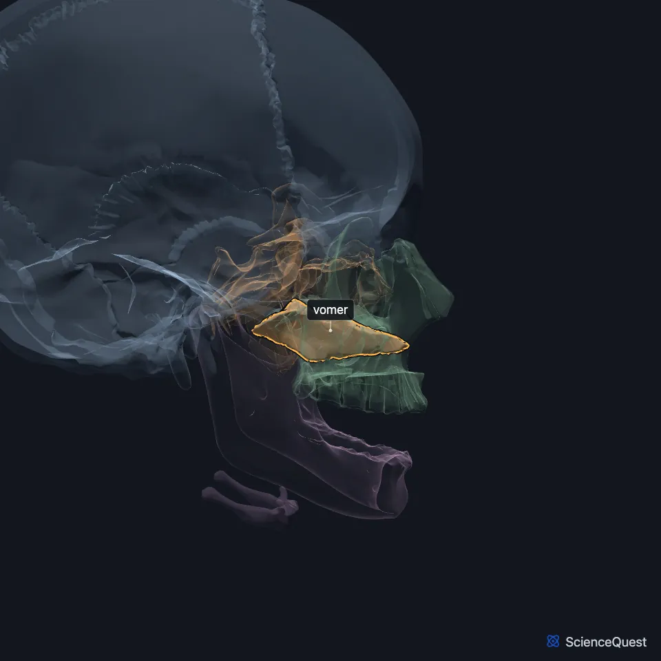
Where is the vomer?
It sits below and slightly behind the perpendicular plate of the ethmoid. Its lower edge rests on the nasal crest that the maxillae and the palatine bones form. (StatPearls, Nasal Cavity; Gray 1918, 5b. 7. The Vomer)
Its back edge is free and concave, and it separates the two choanae. These are the openings at the back of the nasal cavity that lead into the nasopharynx. (Gray 1918, 5b. 7. The Vomer; StatPearls, Nasal Cavity)
What shape is the vomer?
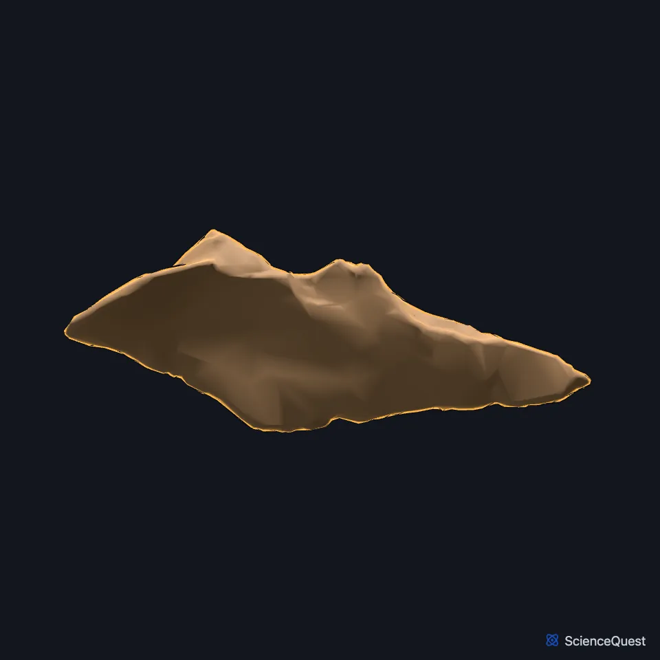
It is a thin, roughly four-sided plate with two surfaces and four borders. On each surface a nasopalatine groove runs down and forward and carries the nasopalatine nerve and vessels. (Gray 1918, 5b. 7. The Vomer)
The upper border is the thickest. A deep furrow there takes the rostrum of the sphenoid, and a wing of bone on each side, the ala, meets the sphenoid behind and the palatine bones in front. (Gray 1918, 5b. 7. The Vomer)
What does the vomer do?
The nasal septum is bone at the back and cartilage at the front. The vomer forms the lower part of the bony septum and the perpendicular plate of the ethmoid the upper part. (StatPearls, Nasal Fractures; Gray 1918, 5b. 7. The Vomer)
Its long front border holds the cartilage in place. The upper half of that border is fused with the perpendicular plate of the ethmoid, and the lower half is grooved for the lower margin of the septal cartilage. (Gray 1918, 5b. 7. The Vomer; StatPearls, Nasal Cavity)
Which bones meet the vomer?
Gray’s Anatomy (1918) counts six: the sphenoid and the ethmoid of the cranium, and both maxillae and both palatine bones of the face. The vomer also joins the septal cartilage of the nose. (Gray 1918, 5b. 7. The Vomer)
How does the vomer form?
Early on, the nasal septum is a plate of cartilage. The vomer ossifies in the membrane over its lower back part, from two centers that appear about the eighth week of fetal life, one on each side of the midline. (Gray 1918, 5b. 7. The Vomer)
So the bone starts as two thin plates, which have almost fully joined by puberty. The flared alae on its upper border and the groove on its front edge are traces of that start. (Gray 1918, 5b. 7. The Vomer)
Is the vomer always straight?
No. Gray’s Anatomy (1918) places the vomer in the midline but notes that its front part is often bent to one side or the other. (Gray 1918, 5b. 7. The Vomer)
| Feature | Detail and source |
|---|---|
| Forms | The lower and back part of the nasal septum (Gray 1918, 5b. 7. The Vomer; StatPearls, Nasal Fractures) |
| Articulates with | 6 bones: sphenoid, ethmoid, both maxillae, both palatine bones; also the septal cartilage (Gray 1918, 5b. 7. The Vomer) |
| Carries | The nasopalatine nerve and vessels, in a groove on each side (Gray 1918, 5b. 7. The Vomer) |
Touches in this model
Measured on the BodyParts3D meshes: surfaces within 2 mm of the vomer, nearest first.
- Ethmoid in contact
- Left palatine bone in contact
- Right palatine bone in contact
- Skin in contact
- Right inferior nasal concha 0.6 mm away
Sources
Sources read
- Gray’s Anatomy of the Human Body, 20th US edition (1918), II. Osteology, 5b. 7. The Vomer. Henry Gray. Public domain. Read .
- StatPearls, Anatomy, Head and Neck, Skull. Bradley W. Anderson; Michael W. Kortz; Asa C. Black; Khalid A. Al Kharazi. Last update . CC BY-NC-ND 4.0. Read .
- StatPearls, Anatomy, Head and Neck, Nasal Cavity. John L. Sobiesk; Sunil Munakomi. Last update . CC BY-NC-ND 4.0. Read .
- StatPearls, Nasal Fractures. Michael Klinginsmith; Marc H. Hohman; Ziad Katrib. Last update . CC BY-NC-ND 4.0. Read .
The values are facts and free to use; this page’s selection and presentation are © 2026 ScienceQuest.