External intercostal muscle
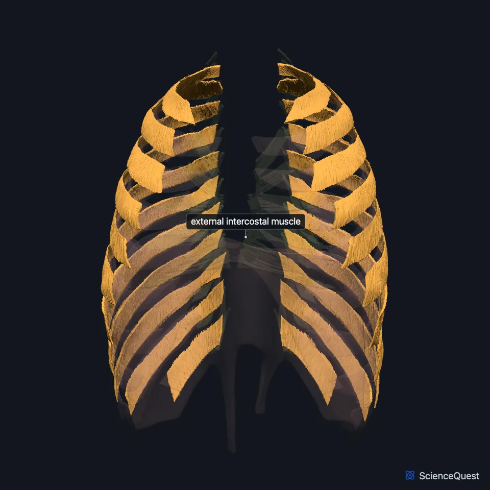
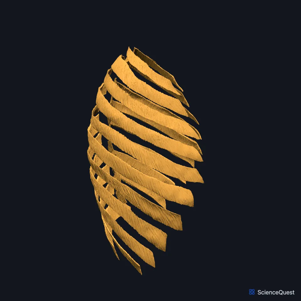
The external intercostal muscles are the outermost of the three muscle layers in each space between the ribs. The 11 pairs help you breathe in: when they contract, they raise and expand the rib cage. (OpenStax A&P 2e, 11.4; StatPearls, Thorax, Muscles)
Open it in the Chest Wall Muscles Explorer See it alone in 3D
Where are the external intercostal muscles?
Eleven pairs of intercostal muscles fill the intercostal spaces in layers from superficial to deep, and the external layer is the outermost. (StatPearls, Thorax, Muscles)
Each runs from the tubercle of the rib at the back to the costochondral junction at the front. There it gives way to a thin sheet of connective tissue, the external intercostal membrane. (StatPearls, Thorax, Muscles)
What do the external intercostals attach to?
Each one arises from the lower border of the rib above and inserts on the upper border of the rib below. Its fibers run down and toward the midline. (StatPearls, Thorax, Muscles)
What do the external intercostals do?
In inspiration they lift the side portions of the ribs and widen the chest from side to side, in what StatPearls calls a bucket-handle motion. (StatPearls, Thorax, Muscles)
In quiet breathing they and the diaphragm are the two muscle groups that draw air in. As the ribs move up the thoracic cavity grows, the pressure in the pleural space falls and the lungs expand. (OpenStax A&P 2e, 22.3; StatPearls, Physiology, Respiratory Drive)
Neuron groups in the medulla oblongata and pons set the baseline breathing rhythm. (OpenStax A&P 2e, 22.3; StatPearls, Physiology, Respiratory Drive)
What lies around the external intercostals?
Under them lie the internal intercostal muscles, whose fibers run at right angles to theirs, and under those the innermost intercostals. (StatPearls, Thorax, Muscles)
Going inward at the side of the chest, StatPearls lists the layers in order: skin, superficial fascia, serratus anterior, the external, internal and innermost intercostals, then the parietal pleura. (StatPearls, Thorax, Muscles)
Intercostal nerves from the anterior rami of T1 to T11 supply the muscles of the thoracic wall. (StatPearls, Thorax, Muscles)
The upper nine spaces each have one posterior and two anterior intercostal arteries: StatPearls traces the anterior arteries of the first six spaces to the internal thoracic artery and those of the seventh to ninth to the musculophrenic artery. From the third space down, the posterior arteries branch straight from the descending thoracic aorta. (StatPearls, Thorax, Muscles)
Where the sources differ
StatPearls’ thorax muscles article has the external intercostals lifting the side portions of the ribs, widening the chest from side to side in a bucket-handle motion. Its diaphragm article has them raising the front of the chest wall like the handles of a bucket. (StatPearls, Thorax, Muscles and Thorax: Diaphragm)
The sources split the breathing rhythm work differently. OpenStax has the ventral respiratory group drive the diaphragm and external intercostals, while StatPearls calls the dorsal group mainly inspiratory and the ventral group mainly expiratory, with rhythm-generating neurons in the ventral group’s rostral half. (OpenStax A&P 2e, 22.3; StatPearls, Physiology, Respiratory Drive)
| Feature | Detail and source |
|---|---|
| Origin | Lower border of the rib above (StatPearls, Thorax, Muscles) |
| Insertion | Upper border of the rib below (StatPearls, Thorax, Muscles) |
| Action | Raise the ribs and widen the chest in inspiration (StatPearls, Thorax, Muscles) |
| Nerve | Intercostal nerves (anterior rami of T1 to T11) (StatPearls, Thorax, Muscles) |
Touches in this model
Measured on the BodyParts3D meshes: surfaces within 2 mm of the external intercostal muscle, nearest first, 12 of 84.
- Diaphragm in contact
- Internal intercostal muscle in contact
- Set of left levatores costarum breves in contact
- Set of left levatores costarum longi in contact
- Set of right levatores costarum breves in contact
- Set of right levatores costarum longi in contact
- Left eighth rib in contact
- Left eleventh rib in contact
- Left external oblique in contact
- Left fifth rib in contact
- Left first rib in contact
- Left fourth rib in contact
Sources
- OpenStax Anatomy and Physiology 2e, section 11.4, Axial Muscles of the Abdominal Wall, and Thorax. CC BY-NC-SA 4.0. Read 2026-10-25.
- StatPearls, Anatomy, Thorax, Muscles. Alex Tang; Bruno Bordoni. Last update July 24, 2023. CC BY-NC-ND 4.0. Read 2026-10-25.
- OpenStax Anatomy and Physiology 2e, section 22.3, The Process of Breathing. CC BY-NC-SA 4.0. Read 2026-10-25.
- StatPearls, Physiology, Respiratory Drive. Joshua E. Brinkman; Fadi Toro; Sandeep Sharma. Last update June 5, 2023. CC BY-NC-ND 4.0. Read 2026-10-25.
- StatPearls, Anatomy, Thorax: Diaphragm. Kanwal Naveen S. Bains; Sarang Kashyap; Sarah L. Lappin. Last update July 24, 2023. CC BY-NC-ND 4.0. Read 2026-10-25.
The values are facts and free to use; this page’s selection and presentation are © 2026 ScienceQuest.