Costal cartilage
Costal cartilages are bars of hyaline cartilage that carry the ribs forward. With the ribs and the sternum they make up the chest wall, and they give it much of its elasticity. (Gray 1918, 4c. The Costal Cartilages; StatPearls, Costochondritis)
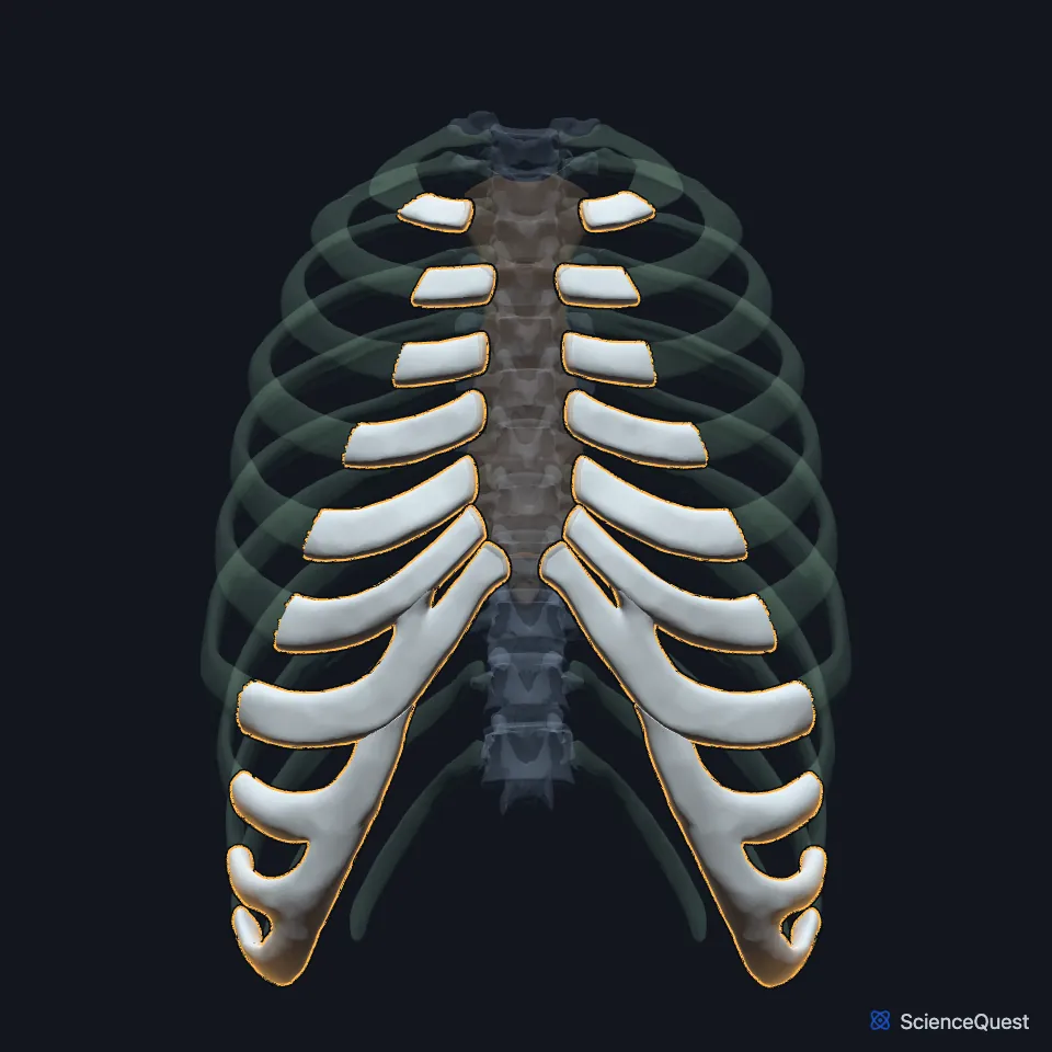
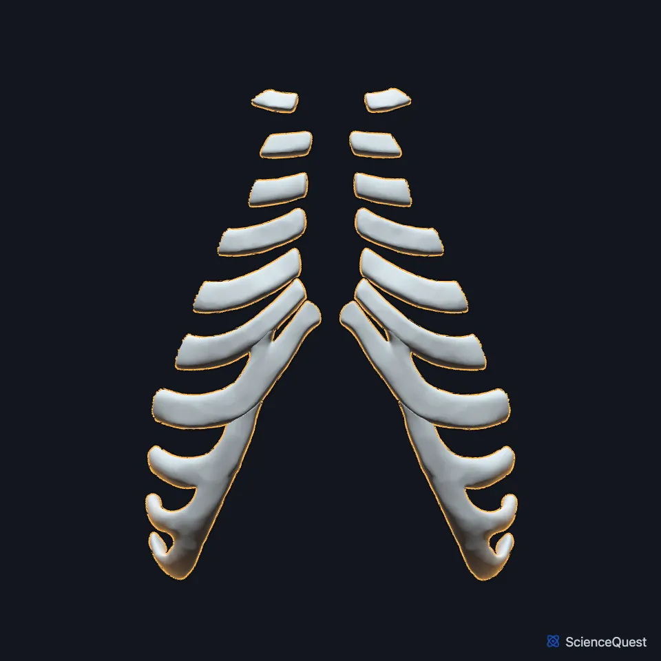
Open it in the Rib Cage and Thoracic Spine Explorer See it alone in 3D
Where are the costal cartilages?
Each one is continuous with the front end of its rib. With the sternum, they form the front boundary of the thoracic wall. (Gray 1918, 4c. The Costal Cartilages; StatPearls, Thorax, Wall)
The first pair joins the manubrium and the second meets the sternum at the sternal angle. The third to seventh pairs join the side of the sternal body, below that angle. (StatPearls, Sternum)
Which ribs reach the sternum?
Ribs 1 to 7 are the true ribs, and each reaches the sternum through its own cartilage. In Gray’s Anatomy (1918) the cartilages of ribs 8 to 10 join the cartilage above instead, and ribs 11 and 12 have pointed, free ends with no sternal attachment. (StatPearls, Thorax, Ribs and Costochondritis; Gray 1918, 4c. The Costal Cartilages)
They lengthen from the first to the seventh and then shorten to the twelfth, and they narrow from first to last. Heel-like projections on the sixth to ninth carry small facets that meet the cartilage below. (Gray 1918, 4c. The Costal Cartilages)
What do the costal cartilages do?
They let the rib cage give. The cage protects the organs of the chest while still allowing the movements of breathing, and the cartilages add much of the springiness of its walls. (StatPearls, Costochondritis; Gray 1918, 4c. The Costal Cartilages)
They also anchor breathing muscles. The costal part of the diaphragm arises from the inner surfaces of the lower six cartilages, and the transversus thoracis inserts on the inner surfaces of the second to sixth and helps with forced breathing out. (StatPearls, Thorax, Ribs; Gray 1918, 6c. Muscles of the Thorax)
What attaches to the costal cartilages?
On the front, the pectoralis major arises from the sternal ends of the first six or seven, and the rectus abdominis inserts on the fifth to seventh. The first gives attachment to the subclavius. (Gray 1918, 4c. The Costal Cartilages; StatPearls, Thorax, Ribs)
Their upper and lower borders give attachment to the internal intercostal muscles. The lower six or seven also give attachment, on their inner surfaces, to the transversus abdominis. (Gray 1918, 4c. The Costal Cartilages)
How do the costal cartilages vary and change?
The costal margin often differs from the textbook pattern. In a 2023 study of 30 cadavers cited by StatPearls, the 10th rib did not join the ninth, and so floated, in more than 50%, and the eighth rib reached the sternum directly in 10%. A cadaver study of the intercostal nerves, cited in the same article, found that direct attachment in 42% of specimens. (StatPearls, Thorax, Ribs)
In old age the cartilages tend to ossify at the surface. Inflammation where they meet the sternum, costochondritis, causes chest wall pain and can mimic a heart attack. It often involves the second to fifth junctions, though any junction can be affected. (Gray 1918, 4c. The Costal Cartilages; StatPearls, Costochondritis)
Where the sources differ
The sources differ on how ribs 8 to 10 reach the sternum. Gray’s Anatomy (1918) joins each of their cartilages to the one directly above, while the StatPearls rib article has all three join the seventh costal cartilage. (Gray 1918, 4c. The Costal Cartilages; StatPearls, Thorax, Ribs)
The sources differ on which cartilages the transversus thoracis reaches. StatPearls and the muscle chapter of Gray’s Anatomy (1918) give the second to sixth, while the costal cartilage section of the same edition gives the third to sixth. (StatPearls, Thorax, Ribs; Gray 1918, 6c. Muscles of the Thorax and 4c. The Costal Cartilages)
| Feature | Detail and source |
|---|---|
| Tissue | Hyaline cartilage (Gray 1918, 4c. The Costal Cartilages) |
| Joins | Ribs 1 to 7: the sternum. Ribs 8 to 10: the cartilage above. Ribs 11 and 12: free (Gray 1918, 4c. The Costal Cartilages) |
Touches in this model
Measured on the BodyParts3D meshes: surfaces within 2 mm of the costal cartilage, nearest first, 12 of 58.
- Body of sternum in contact
- Left eighth rib in contact
- Left fifth rib in contact
- Left first rib in contact
- Left fourth rib in contact
- Left ninth rib in contact
- Left second rib in contact
- Left seventh rib in contact
- Left sixth rib in contact
- Left tenth rib in contact
- Left third rib in contact
- Manubrium in contact
Sources
Sources read
- Gray’s Anatomy of the Human Body, 20th US edition (1918), II. Osteology, 4c. The Costal Cartilages. Henry Gray. Public domain. Read .
- StatPearls, Costochondritis. Jessica A. Schumann; Tanuj Sood; John J. Parente. Last update . CC BY-NC-ND 4.0. Read .
- StatPearls, Anatomy, Thorax, Wall. Azhar Hussain; Bracken Burns. Last update . CC BY-NC-ND 4.0. Read .
- StatPearls, Anatomy, Thorax, Sternum. Abdulraheem A. Altalib; Kathleen H. Miao; Ritesh G. Menezes. Last update . CC BY-NC-ND 4.0. Read .
- StatPearls, Anatomy, Thorax, Ribs. Omar A. Safarini; Bruno Bordoni. Last update . CC BY-NC-ND 4.0. Read .
- Gray’s Anatomy of the Human Body, 20th US edition (1918), IV. Myology, 6c. The Muscles of the Thorax (1918 terms: Intercostales externi and interni for the external and internal intercostals, anterior intercostal membranes for external intercostal membranes). Henry Gray. Public domain. Read .
The values are facts and free to use; this page’s selection and presentation are © 2026 ScienceQuest.