Thoracic vertebrae
The thoracic vertebrae are the 12 vertebrae of the chest, T1 to T12, between the cervical and lumbar vertebrae. Their costal facets, the joint surfaces for the ribs, set them apart. (StatPearls, Thoracic Vertebrae; Gray’s Anatomy (1918), Thoracic vertebrae, p. 102)
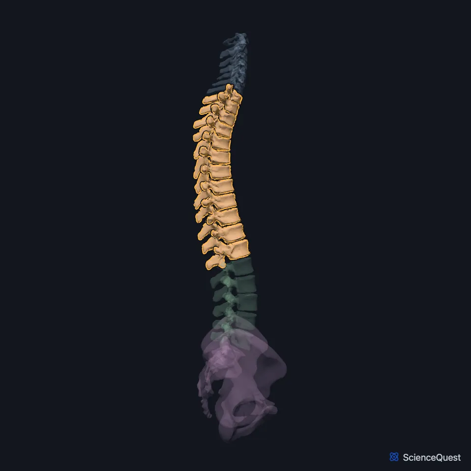
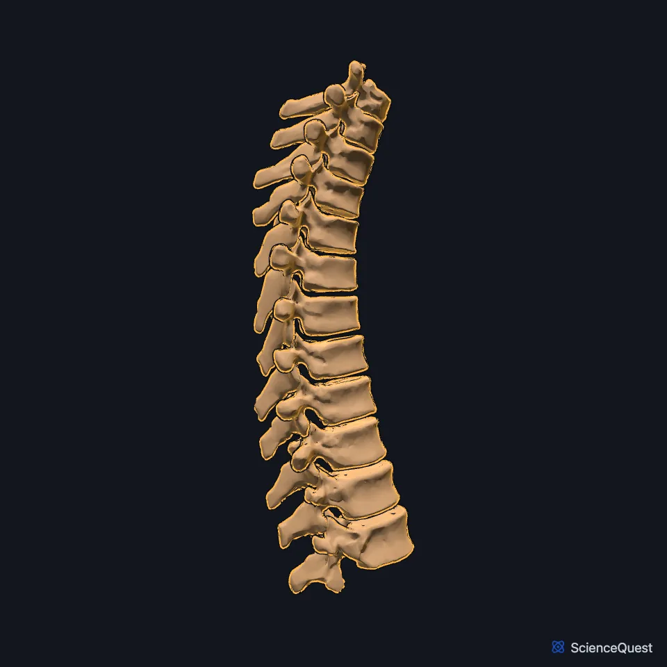
Open it in the Vertebral Column and Pelvis Explorer See it alone in 3D
Where are the thoracic vertebrae?
They form the part of the column between the neck and the lower back, and together they curve the spine backwards in a kyphosis. They grow larger from T1 down to T12. (StatPearls, Thoracic Vertebrae; Gray’s Anatomy (1918), Thoracic vertebrae, p. 102)
How do the ribs attach to the thoracic vertebrae?
A typical thoracic vertebra has six costal facets: a half facet above and below on each side of the body, and one on each transverse process. The head of a rib sits across two vertebrae, so the lower half facet of T4 and the upper half facet of T5 both take the head of rib 5, and the facet on the transverse process takes the rib’s tubercle. (StatPearls, Thoracic Vertebrae)
The half facets and the intervertebral disc between them make one oval socket for each rib head. (Gray’s Anatomy (1918), Thoracic vertebrae, p. 102)
What does a thoracic vertebra look like?
In the middle of the region the bodies are heart-shaped. The laminae overlap the ones below like tiles on a roof, and the long spinous processes slope downward, overlapping from the fifth to the eighth. (Gray’s Anatomy (1918), Thoracic vertebrae, p. 102; StatPearls, Thoracic Vertebrae)
What do the thoracic vertebrae do?
They carry the ribs and, with them, support the thorax, and their arches enclose the spinal cord. The ribs and the shape of the spinous processes limit bending forward and back here, and T5 to T8 rotate the most. (StatPearls, Thoracic Vertebrae)
The thoracic canal is narrow, which makes the cord more open to injury. Spinal injuries cluster at the junction with the lumbar spine, where the rigid thoracic column meets the more mobile lumbar one. (StatPearls, Thoracic Vertebrae)
Which thoracic vertebrae are atypical?
T1 has a whole facet for the first rib and a half facet for the second. T11 and T12 each carry one whole facet for their own rib and none on the transverse processes. (StatPearls, Thoracic Vertebrae; Gray’s Anatomy (1918), Thoracic vertebrae, p. 102)
Where the sources differ
StatPearls counts three atypical thoracic vertebrae, T1, T11 and T12, with T10 sometimes like them. Gray’s Anatomy (1918) singles out five, the first, ninth, tenth, eleventh and twelfth. (StatPearls, Thoracic Vertebrae; Gray’s Anatomy (1918), Thoracic vertebrae, p. 102)
| Feature | Detail and source |
|---|---|
| Number | 12, T1 to T12 (StatPearls, Thoracic Vertebrae) |
| Articulate with | Ribs (heads and tubercles); the vertebrae above and below (StatPearls, Thoracic Vertebrae) |
| Curve | Kyphosis (StatPearls, Thoracic Vertebrae) |
Touches in this model
Measured on the BodyParts3D meshes: surfaces within 2 mm of the thoracic vertebrae, nearest first, 12 of 102.
- Seventh cervical vertebra in contact
- Ascending part of right trapezius in contact
- Descending aorta in contact
- Diaphragm in contact
- Esophagus in contact
- External intercostal muscle in contact
- Inferior oblique part of right longus colli in contact
- Intervertebral disk of eighth thoracic vertebra in contact
- Intervertebral disk of eleventh thoracic vertebra in contact
- Intervertebral disk of fifth thoracic vertebra in contact
- Intervertebral disk of first thoracic vertebra in contact
- Intervertebral disk of fourth thoracic vertebra in contact
Sources
Sources read
- StatPearls, Anatomy, Back, Thoracic Vertebrae. Joshua A. Waxenbaum; Vamsi Reddy; Konstantinos Margetis. Last update . CC BY-NC-ND 4.0. Read .
- Gray’s Anatomy of the Human Body, 20th US edition (1918), edited by Warren H. Lewis, Osteology, The Thoracic Vertebrae (pp. 102 to 104); 1918 name: intervertebral fibrocartilages for intervertebral disks. Henry Gray; Warren H. Lewis (editor). Public domain. Read .
The values are facts and free to use; this page’s selection and presentation are © 2026 ScienceQuest.