Longus colli
The longus colli lies on the front of the vertebral column, from the atlas to the third thoracic vertebra. It is broad in the middle, narrow at each end, and has three parts. (Gray 1918, 5d. Anterior Vertebral Muscles; StatPearls, Prevertebral Muscles)
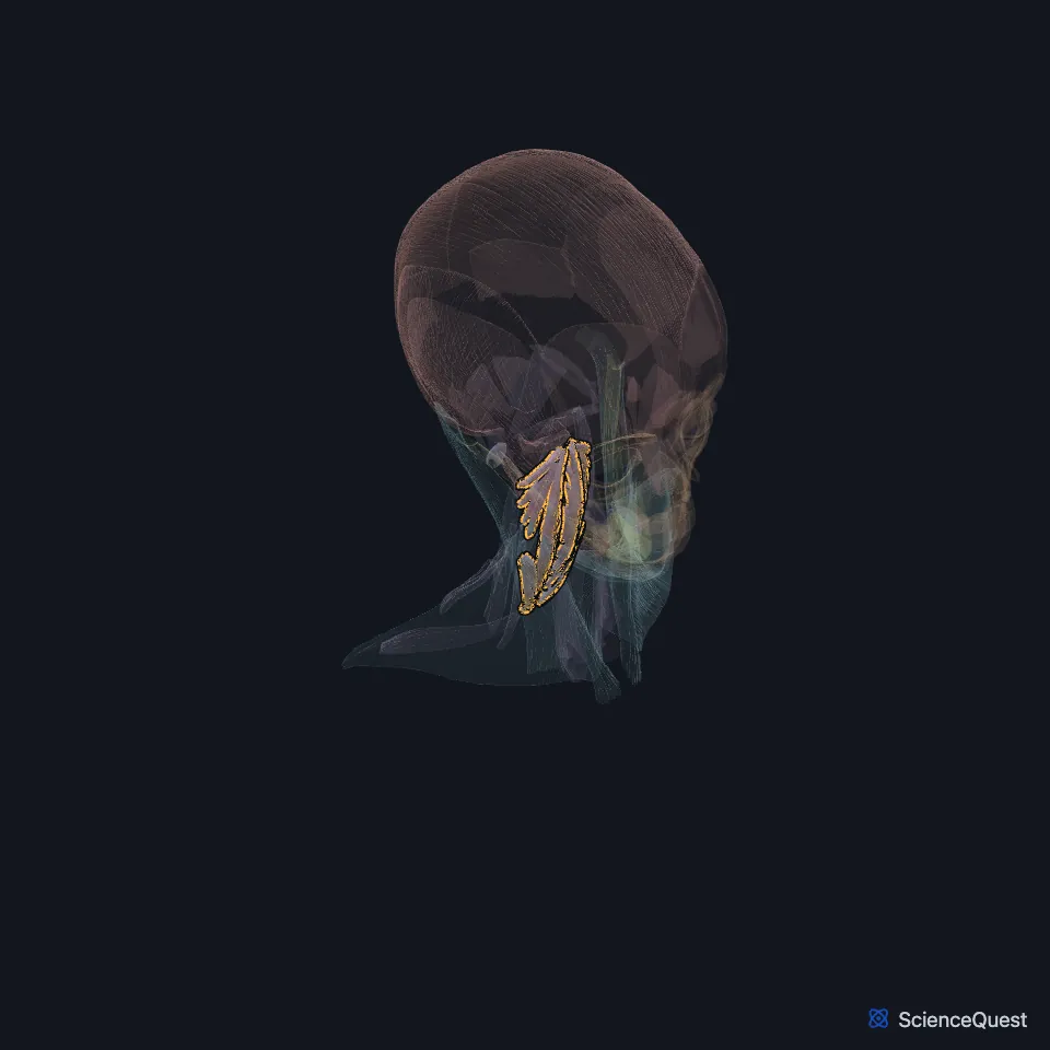
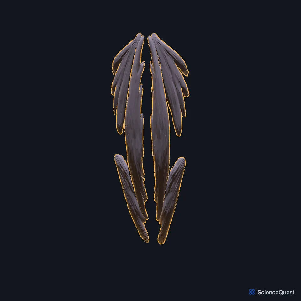
Open it in the Head and Neck Muscles Explorer See it alone in 3D
Where is the longus colli?
It is one of four prevertebral muscles, with the longus capitis, the rectus capitis anterior and the rectus capitis lateralis. They lie behind the prevertebral fascia and in front of the cervical spine. (StatPearls, Prevertebral Muscles)
What are the three parts of the longus colli?
The superior oblique part runs from the anterior tubercles of the C3 to C5 transverse processes up and inward to the tubercle on the front of the atlas. (Gray 1918, 5d. Anterior Vertebral Muscles)
The inferior oblique part, the smallest, runs from the front of the bodies of the first two or three thoracic vertebrae up and outward to the anterior tubercles of C5 and C6. (Gray 1918, 5d. Anterior Vertebral Muscles)
The vertical part runs from the front of the bodies of the upper three thoracic and lower three cervical vertebrae to the bodies of C2 to C4. (Gray 1918, 5d. Anterior Vertebral Muscles)
What does the longus colli do?
Gray’s 1918 text gives its action as flexing the cervical spine and turning it a little. (Gray 1918, 5d. Anterior Vertebral Muscles)
What lies in front of the longus colli?
In front of it lie the prevertebral fascia, the pharynx, the esophagus, the cervical sympathetic trunk, the carotid sheath, the inferior thyroid artery and the recurrent laryngeal nerve. Behind it is the cervical spine. (StatPearls, Prevertebral Muscles)
Spine surgeons use its medial border as a landmark to expose the cervical disc spaces without injuring the sympathetic trunk. (StatPearls, Prevertebral Muscles)
Its blood comes from the ascending cervical branch of the inferior thyroid artery, the ascending pharyngeal artery and the vertebral artery. (StatPearls, Prevertebral Muscles)
What is longus colli tendinitis?
Calcium deposits in its tendon can set off an inflammation called acute calcific tendinitis. It brings sudden neck pain or stiffness, trouble swallowing and sometimes fever, and doctors can mistake it for a retropharyngeal abscess. (StatPearls, Prevertebral Muscles)
Where the sources differ
StatPearls gives its nerves as branches of the C2 to C6 ventral rami. Gray’s 1918 text gives branches of the second to seventh cervical nerves. (StatPearls, Prevertebral Muscles; Gray 1918, 5d. Anterior Vertebral Muscles)
For the superior oblique part, StatPearls gives an insertion on the transverse process and anterior tubercle of C1. Gray’s 1918 text gives the tubercle on the anterior arch of the atlas. (StatPearls, Prevertebral Muscles; Gray 1918, 5d. Anterior Vertebral Muscles)
StatPearls has the two longus colli muscles flexing the head together and one alone tilting it at the craniocervical junction. Gray’s 1918 text limits its action to the cervical spine, which it flexes and turns slightly, and in the same passage credits head flexion to the longus capitis and rectus capitis anterior. (StatPearls, Prevertebral Muscles; Gray 1918, 5d. Anterior Vertebral Muscles)
| Feature | Detail and source |
|---|---|
| Origin | Superior oblique: C3 to C5 transverse processes. Inferior oblique: bodies of T1 to T2 or T3. Vertical: bodies of the lower three cervical and upper three thoracic vertebrae (Gray 1918, 5d. Anterior Vertebral Muscles) |
| Insertion | Superior oblique: anterior arch of the atlas. Inferior oblique: C5 and C6 transverse processes. Vertical: bodies of C2 to C4 (Gray 1918, 5d. Anterior Vertebral Muscles) |
| Action | Flexes the cervical spine and turns it slightly (Gray 1918, 5d. Anterior Vertebral Muscles) |
| Nerve | Cervical ventral rami: C2 to C6 in StatPearls, C2 to C7 in Gray 1918 (StatPearls, Prevertebral Muscles; Gray 1918, 5d. Anterior Vertebral Muscles) |
Touches in this model
Measured on the BodyParts3D meshes: surfaces within 2 mm of the longus colli, nearest first, 12 of 33.
- Left longus capitis in contact
- Left scalenus anterior in contact
- Right longus capitis in contact
- Right scalenus anterior in contact
- Atlas in contact
- Axis in contact
- Esophagus in contact
- Fifth cervical vertebra in contact
- First thoracic vertebra in contact
- Fourth cervical vertebra in contact
- Intervertebral disk of fifth cervical vertebra in contact
- Intervertebral disk of first thoracic vertebra in contact
Sources
Sources read
- Gray’s Anatomy of the Human Body, 20th US edition (1918), IV. Myology, 5d. The Anterior Vertebral Muscles (1918 terms: Rectus capitis anticus major for longus capitis, Rectus capitis anticus minor for rectus capitis anterior). Henry Gray. Public domain. Read .
- StatPearls, Anatomy, Head and Neck, Prevertebral Muscles. Laura J. McDavid; Yusuf S. Khan. Last update . CC BY-NC-ND 4.0. Read .
The values are facts and free to use; this page’s selection and presentation are © 2026 ScienceQuest.