Scalenus anterior
The scalenus anterior is the front one of the three scalene muscles. It lies deep at the side of the neck, behind the sternocleidomastoid, and runs from the cervical vertebrae down to the first rib. (Gray 1918, 5e. Lateral Vertebral Muscles; StatPearls, Scalenus Muscle)
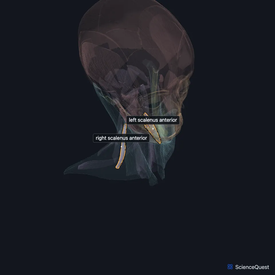
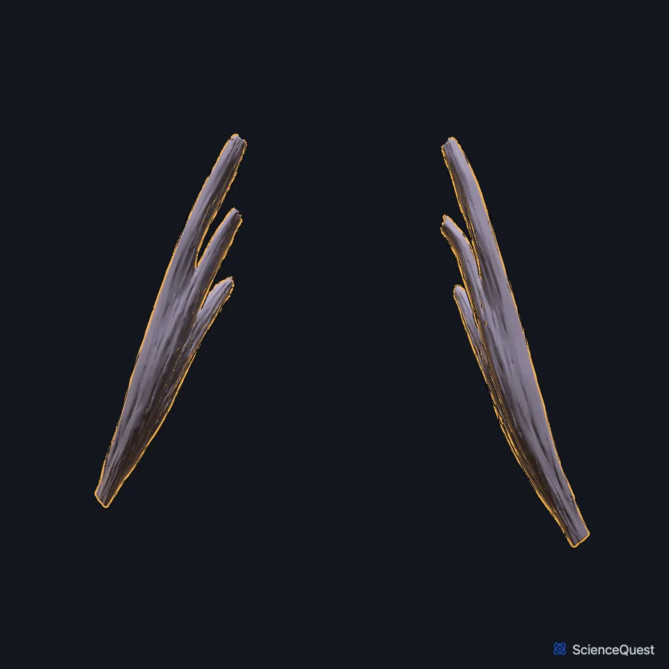
Open it in the Head and Neck Muscles Explorer See it alone in 3D
Where is the scalenus anterior?
It arises from the anterior tubercles of the transverse processes of the third to sixth cervical vertebrae. It descends almost vertically to the scalene tubercle on the inner border and upper surface of the first rib. (Gray 1918, 5e. Lateral Vertebral Muscles; StatPearls, Scalenus Muscle)
It is thickest at the level of the cricoid cartilage and thinner at both ends, and some of its fibers may touch the dome of the pleura. (StatPearls, Scalenus Muscle)
What does the scalenus anterior do?
As a group, the scalenes lift the first and second ribs, so they work as accessory muscles of breathing in forced inspiration. StatPearls notes that their electrical activity continues even in quiet breathing. (Gray 1918, 5e. Lateral Vertebral Muscles; StatPearls, Scalenus Muscle)
With the ribs as the fixed end, the scalenes bend the neck to one side, and both sides together flex it a little. They also help hold the neck’s posture. (Gray 1918, 5e. Lateral Vertebral Muscles; StatPearls, Interscalene Triangle)
What runs beside the scalenus anterior?
The phrenic nerve, which drives the diaphragm, runs down across its front surface inside the muscle’s fascia. (StatPearls, Scalenus Muscle and Thorax, Phrenic Nerves)
Its lateral border, the scalenus medius and the first rib frame the interscalene triangle. The roots and trunks of the brachial plexus and the subclavian artery pass through that gap, while the subclavian vein stays outside it. (StatPearls, Interscalene Triangle)
The thyrocervical trunk rises along its medial side, and the trunk’s inferior thyroid branch supplies the muscle. (StatPearls, Scalenus Muscle)
Why does the scalenus anterior matter in thoracic outlet syndrome?
Thoracic outlet syndrome is compression of the subclavian vessels or the brachial plexus between the clavicle, the scalenes and the first rib. To relieve it, surgeons may remove part or all of the scalenus anterior. (StatPearls, Scalenus Muscle)
How does the scalenus anterior vary?
Its origin may start at C2 or skip C6, and its insertion may reach the second or third rib. A slip of it may pass behind the subclavian artery, and in rare cases the muscle is missing on one or both sides. (StatPearls, Scalenus Muscle; Gray 1918, 5e. Lateral Vertebral Muscles)
A fourth scalene, the scalenus minimus, is present in 30% to 71% of people. It runs from the transverse process of C7 to the first rib and the fascia over the dome of the pleura; Gray’s 1918 text calls a muscle with these attachments the scalenus pleuralis. (StatPearls, Scalenus Muscle; Gray 1918, 5e. Lateral Vertebral Muscles)
Where the sources differ
StatPearls gives the scalenes’ nerve supply as the anterior branches of C3 to C8, through both the cervical and brachial plexuses. Gray’s 1918 text gives branches of the second to seventh cervical nerves. (StatPearls, Scalenus Muscle; Gray 1918, 5e. Lateral Vertebral Muscles)
StatPearls’ scalene article puts cervical ribs at about 0.5% of people. Its interscalene triangle article gives about 2%, and nearly 30% among people with thoracic outlet syndrome. (StatPearls, Scalenus Muscle and Interscalene Triangle)
| Feature | Detail and source |
|---|---|
| Origin | Anterior tubercles of the transverse processes of C3 to C6 (Gray 1918, 5e. Lateral Vertebral Muscles; StatPearls, Scalenus Muscle) |
| Insertion | Scalene tubercle of the first rib (Gray 1918, 5e. Lateral Vertebral Muscles; StatPearls, Scalenus Muscle) |
| Action | Lifts the first rib in forced inspiration; bends the neck to the side (Gray 1918, 5e. Lateral Vertebral Muscles; StatPearls, Scalenus Muscle) |
| Nerve | Anterior rami of cervical nerves (see the note below on which ones) (StatPearls, Scalenus Muscle; Gray 1918, 5e. Lateral Vertebral Muscles) |
Touches in this model
Measured on the BodyParts3D meshes: surfaces within 2 mm of the scalenus anterior, nearest first, 12 of 22.
- Inferior oblique part of left longus colli in contact
- Inferior oblique part of right longus colli in contact
- Left longus capitis in contact
- Left omohyoid in contact
- Right longus capitis in contact
- Right omohyoid in contact
- Superior oblique part of left longus colli in contact
- Superior oblique part of right longus colli in contact
- Fifth cervical vertebra in contact
- Fourth cervical vertebra in contact
- Left first rib in contact
- Left subclavian artery in contact
Sources
Sources read
- Gray’s Anatomy of the Human Body, 20th US edition (1918), IV. Myology, 5e. The Lateral Vertebral Muscles (1918 terms: Scalenus anticus for scalenus anterior, Scalenus posticus for scalenus posterior). Henry Gray. Public domain. Read .
- StatPearls, Anatomy, Head and Neck, Scalenus Muscle. Bruno Bordoni; Felix Jozsa; Matthew A. Varacallo. Last update . CC BY-NC-ND 4.0. Read .
- StatPearls, Anatomy, Head and Neck: Interscalene Triangle. James La Rose; Felix Jozsa. Last update . CC BY-NC-ND 4.0. Read .
- StatPearls, Anatomy, Thorax, Phrenic Nerves. Kaitlin A. Oliver; John V. Ashurst. Last update . CC BY-NC-ND 4.0. Read .
The values are facts and free to use; this page’s selection and presentation are © 2026 ScienceQuest.