Clavicle
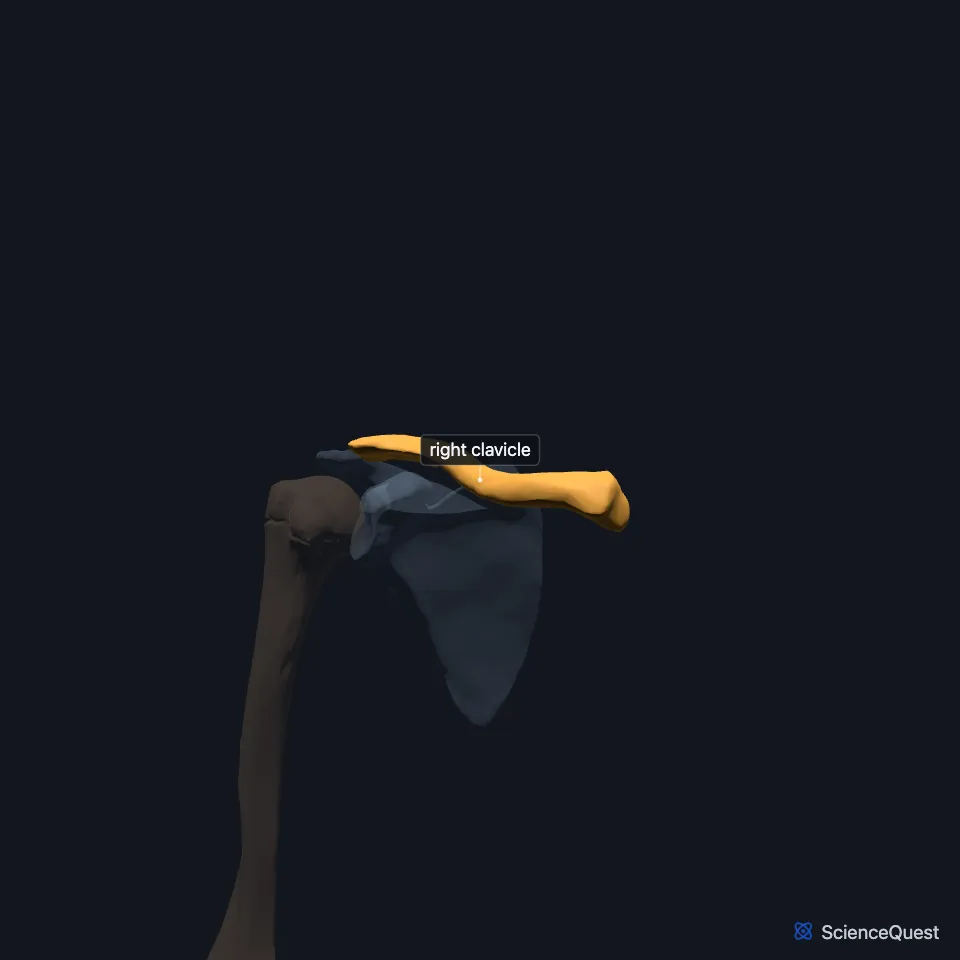
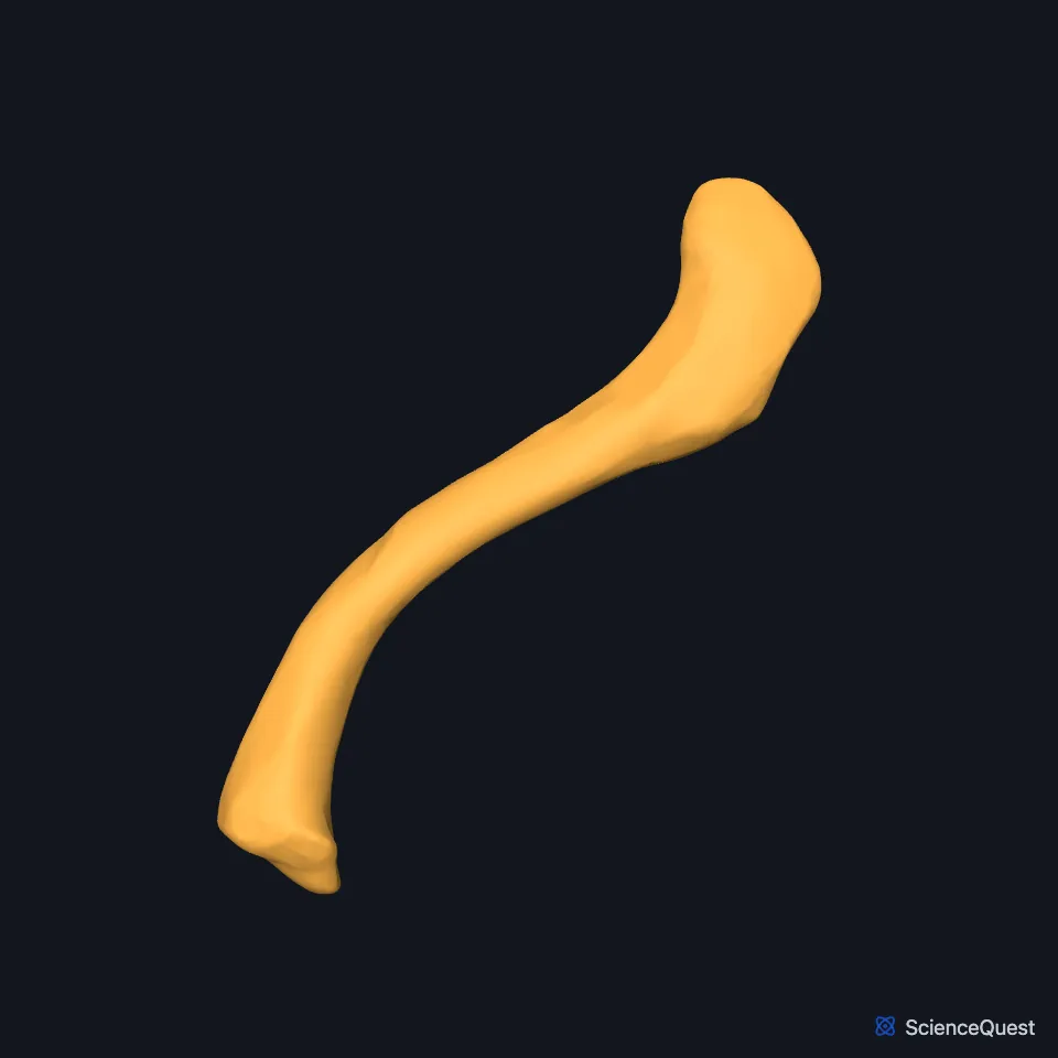
The clavicle, or collarbone, is an S-shaped bone on the front of the shoulder. With the scapula it forms the pectoral girdle, which attaches the upper limb to the axial skeleton. (OpenStax A&P 2e, 8.1; StatPearls, Clavicle and Clavicle Fracture; Gray’s Anatomy (1918), Clavicle, p. 200)
Open it in the Upper Limb Skeleton Explorer See it alone in 3D
Where is the clavicle?
The clavicle lies nearly horizontal across the top of the chest, just above the first rib. It sits just under the skin, so you can feel it. (OpenStax A&P 2e, 8.1; Gray’s Anatomy (1918), Clavicle, p. 200; StatPearls, Clavicle Fracture)
Its medial end attaches to the sternum, part of the axial skeleton. Its lateral end meets the scapula just above the shoulder joint. (OpenStax A&P 2e, 8.1; StatPearls, Clavicle and Sternoclavicular Joint; Gray’s Anatomy (1918), Scapula, p. 202)
What does the clavicle do?
Held from above by muscles, the clavicle acts as a strut that props the scapula out to the side. That keeps the shoulder joint up and away from the trunk and gives the upper limb its freedom of motion. (OpenStax A&P 2e, 8.1; StatPearls, Clavicle)
It passes forces from the upper limb to the sternum and axial skeleton. It also shields the nerves and vessels that run beneath it between the trunk and the limb: StatPearls names the brachial plexus, the subclavian artery and the subclavian vein. (OpenStax A&P 2e, 8.1; StatPearls, Clavicle)
What are the parts of the clavicle?
The clavicle has a medial end, a lateral end and a shaft between them. The medial or sternal end is triangular. The lateral or acromial end meets the acromion, the part of the scapula that forms the tip of the shoulder. (OpenStax A&P 2e, 8.1; Gray’s Anatomy (1918), Clavicle, p. 200; StatPearls, Clavicle and Scapula)
The clavicle starts to ossify before any other bone in the embryo, yet its growth plates may not close until age 20 to 25. Its two ends ossify from a cartilage model; the shaft forms bone with no cartilage first. (StatPearls, Clavicle)
Blood reaches it through the suprascapular, thoracoacromial and internal thoracic arteries. Studies have found a supply through its periosteum and no central nutrient artery. (StatPearls, Clavicle)
Which joints and muscles does the clavicle have?
Its sternal end meets the manubrium of the sternum at the sternoclavicular joint, the only bony joint between the pectoral girdle and the axial skeleton. The joint lets the clavicle and scapula move up and down and forward and back with the shoulder. (OpenStax A&P 2e, 8.1; StatPearls, Shoulder; Gray’s Anatomy (1918), Sternoclavicular joint, p. 313)
The costoclavicular ligament ties the underside of the sternal end to the first rib; OpenStax says it supports the sternoclavicular joint indirectly, StatPearls calls it the joint’s primary restraint, and Gray’s Anatomy (1918) counts it among the joint’s ligaments. At the other end, the acromioclavicular joint with the acromion takes its main support from the coracoclavicular ligament, which ties the coracoid process of the scapula to the underside of the clavicle. (OpenStax A&P 2e, 8.1; StatPearls, Sternoclavicular Joint and Acromioclavicular Joint; Gray’s Anatomy (1918), Acromioclavicular joint, p. 315 and Sternoclavicular joint, p. 313)
On the lateral third, the deltoid attaches in front and the trapezius behind. The sternocleidomastoid attaches to the upper surface of the medial third. On the medial two-thirds, the subclavius sits in the groove beneath, the pectoralis major in front and the sternohyoid behind. (StatPearls, Clavicle)
Why does the clavicle break so often?
A fall onto the side of the shoulder causes most clavicle fractures, 87 percent of reported cases in StatPearls; a direct blow or a fall onto an outstretched hand causes fewer. The middle third breaks in about 69 percent of cases, while injury to the sternoclavicular joint is rare, 3 to 5 percent of shoulder girdle injuries. (OpenStax A&P 2e, 8.1; StatPearls, Clavicle Fracture and Sternoclavicular Joint)
OpenStax says the vessels and nerves under the clavicle seldom suffer in a fracture, because the broken bone moves forward. StatPearls still lists brachial plexus and subclavian vessel injury among the complications, since both lie close to the middle of the bone, and calls for a complete neurovascular examination. (OpenStax A&P 2e, 8.1; StatPearls, Clavicle Fracture)
How does the clavicle differ between people?
Clavicles differ by sex: men’s are usually longer, thicker and more curved than women’s. (OpenStax A&P 2e, 8.1; StatPearls, Clavicle)
Cadaver studies have measured left clavicles longer than right ones. In the rare genetic disorder cleidocranial dysplasia, the clavicles can be missing on both sides or partly missing. (StatPearls, Clavicle)
Where the sources differ
OpenStax calls the clavicle the bone fractured most often in the body. StatPearls calls it one of the bones fractured most often. (OpenStax A&P 2e, 8.1; StatPearls, Clavicle)
| Feature | Detail and source |
|---|---|
| Bone type | Long bone, lying nearly horizontal (OpenStax A&P 2e, 8.1; StatPearls, Clavicle; Gray’s Anatomy (1918), Clavicle, p. 200) |
| Part of | Pectoral (shoulder) girdle, with the scapula (OpenStax A&P 2e, 8.1; StatPearls, Clavicle) |
| Articulates with | Manubrium of the sternum (sternoclavicular joint); acromion of the scapula (acromioclavicular joint) (OpenStax A&P 2e, 8.1; StatPearls, Clavicle) |
| Arteries | Suprascapular, thoracoacromial and internal thoracic (StatPearls, Clavicle) |
Touches in this model
Measured on the BodyParts3D meshes: surfaces within 2 mm of the clavicle, nearest first, 12 of 19.
- Right scapula in contact
- Clavicular part of right deltoid in contact
- Clavicular part of right pectoralis major in contact
- Descending part of right trapezius in contact
- Manubrium in contact
- Right brachiocephalic vein in contact
- Right platysma in contact
- Right sternocleidomastoid in contact
- Right sternohyoid in contact
- Right subclavian artery in contact
- Right subclavian vein in contact
- Right subclavius in contact
Sources
- OpenStax Anatomy and Physiology 2e, section 8.1, The Pectoral Girdle. CC BY-NC-SA 4.0. Read 2026-10-25.
- StatPearls, Anatomy, Shoulder and Upper Limb, Clavicle. James La Rose; Matthew Charlick; Matthew A. Varacallo. Last update July 15, 2026. CC BY-NC-ND 4.0. Read 2026-10-25.
- StatPearls, Clavicle Fracture. Minh Quan T. Le; Keith Myers; Todd May; Shayan Hosseinzadeh. Last update March 27, 2026. CC BY-NC-ND 4.0. Read 2026-10-25.
- Gray’s Anatomy of the Human Body, 20th US edition (1918), edited by Warren H. Lewis, Osteology, The Clavicle (pp. 200 to 202). Henry Gray; Warren H. Lewis (editor). Public domain. Read 2026-10-25.
- StatPearls, Anatomy, Shoulder and Upper Limb, Sternoclavicular Joint. Thomas N. Epperson; Asa C. Black; Matthew A. Varacallo. Last update November 17, 2023. CC BY-NC-ND 4.0. Read 2026-10-25.
- Gray’s Anatomy of the Human Body, 20th US edition (1918), edited by Warren H. Lewis, Osteology, The Scapula (pp. 202 to 209); 1918 names: vertebral border for medial border, axillary border for lateral border, medial angle for superior angle. Henry Gray; Warren H. Lewis (editor). Public domain. Read 2026-10-25.
- StatPearls, Anatomy, Thorax, Scapula. Mohammed A. Miniato; Andrew Mudreac; Judith Borger. Last update July 24, 2023. CC BY-NC-ND 4.0. Read 2026-10-25.
- StatPearls, Anatomy, Shoulder and Upper Limb, Shoulder. Mohammed A. Miniato; Prashanth Anand; Matthew A. Varacallo. Last update July 24, 2023. CC BY-NC-ND 4.0. Read 2026-10-25.
- Gray’s Anatomy of the Human Body, 20th US edition (1918), edited by Warren H. Lewis, Syndesmology, Sternoclavicular Articulation (pp. 313 to 315). Henry Gray; Warren H. Lewis (editor). Public domain. Read 2026-10-25.
- StatPearls, Anatomy, Shoulder and Upper Limb, Acromioclavicular Joint. James La Rose; Vivek Tiwari. Last update May 23, 2026. CC BY-NC-ND 4.0. Read 2026-10-25.
- Gray’s Anatomy of the Human Body, 20th US edition (1918), edited by Warren H. Lewis, Syndesmology, Acromioclavicular Articulation (pp. 315 to 317). Henry Gray; Warren H. Lewis (editor). Public domain. Read 2026-10-25.
The values are facts and free to use; this page’s selection and presentation are © 2026 ScienceQuest.