Humerus
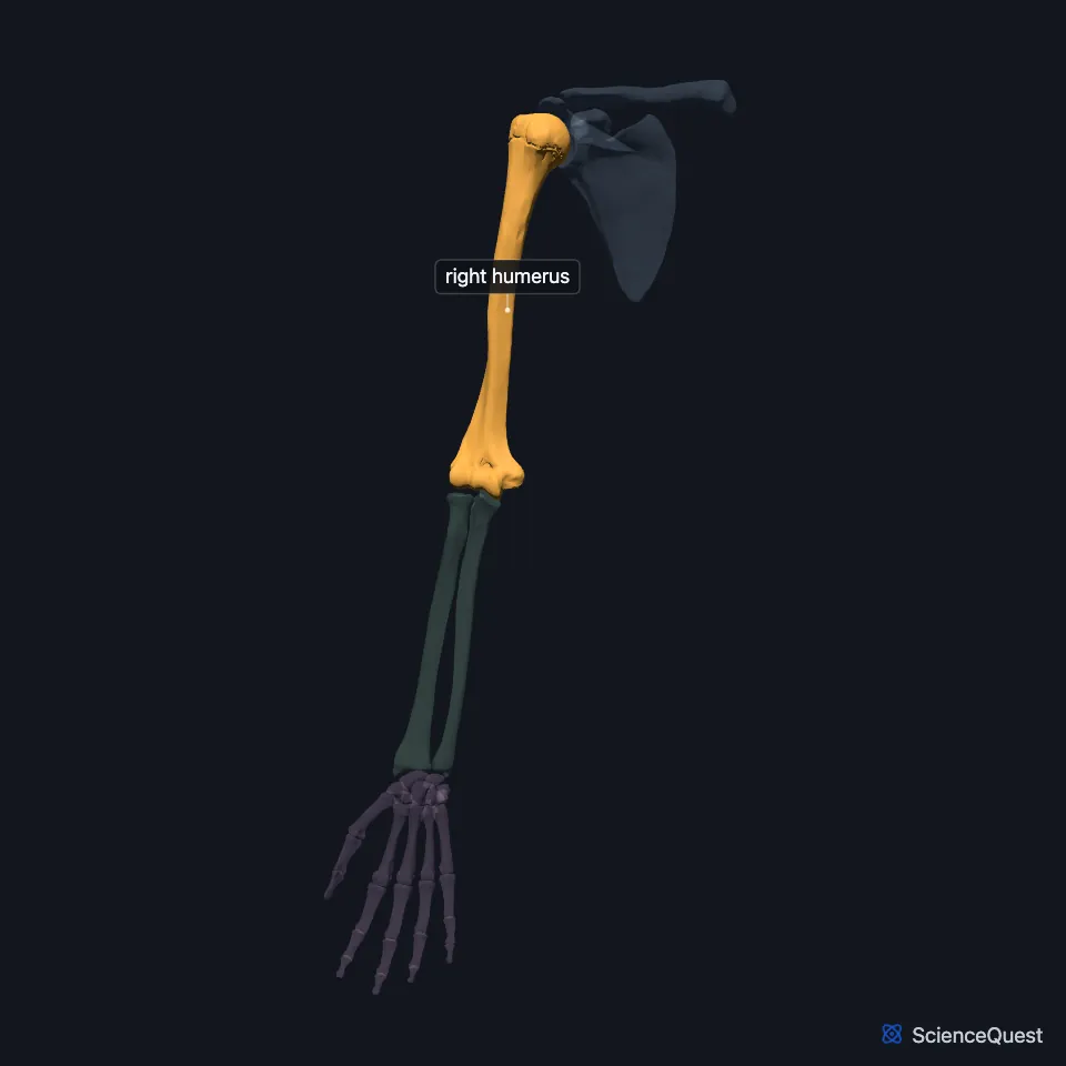
The humerus is the single bone of the arm, between the shoulder and the elbow. StatPearls calls it the largest bone of the upper limb. (OpenStax A&P 2e, 8.2; StatPearls, Humerus)
Open it in the Upper Limb Skeleton Explorer See it alone in 3D
Where is the humerus?
At its upper end the humerus meets the glenoid cavity of the scapula at the shoulder, and at its lower end the radius and ulna at the elbow. (StatPearls, Humerus)
What are the parts of the humerus?
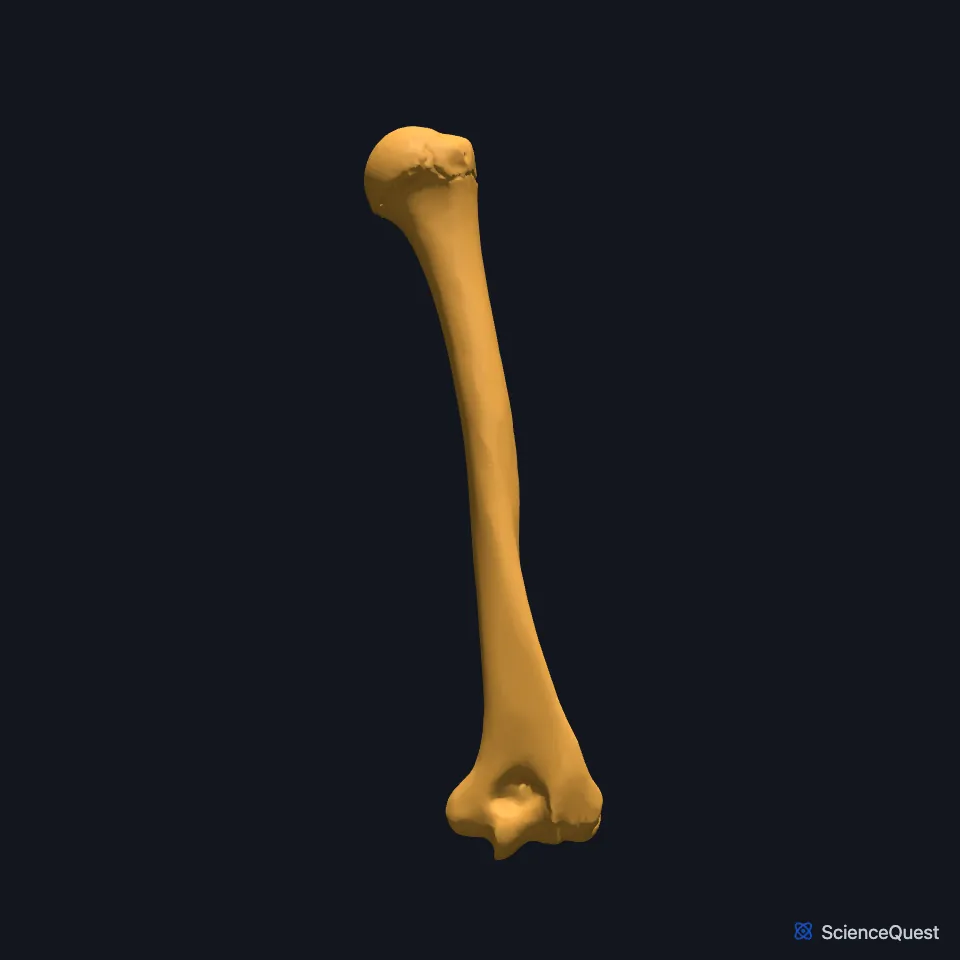
At the top, the large round head faces medially, with the anatomical neck around its margin. The greater tubercle sits on the lateral side and the lesser tubercle on the front, and between them the intertubercular groove carries a tendon of the biceps brachii. (OpenStax A&P 2e, 8.2; Gray’s Anatomy (1918), Humerus, p. 209; StatPearls, Humerus)
The surgical neck, where the wide upper end joins the shaft, is a common site of arm fractures. Halfway down the shaft on the lateral side is the rough deltoid tuberosity, where the deltoid attaches. (OpenStax A&P 2e, 8.2; StatPearls, Humerus and Humerus Fractures Overview; Gray’s Anatomy (1918), Humerus, p. 209)
At the lower end, the medial and lateral epicondyles and the lateral supracondylar ridge above the lateral one anchor muscles that act on the forearm, wrist and hand. The medial epicondyle is the larger, and most of the superficial muscles on the front of the forearm arise from it by a common flexor tendon. (OpenStax A&P 2e, 8.2; Gray’s Anatomy (1918), Humerus, p. 209; StatPearls, Arm Structure and Function)
How does the humerus form the elbow?
The trochlea, a pulley-shaped surface on the medial side of the lower end, meets the ulna. The knob-like capitulum just lateral to it meets the radius. (OpenStax A&P 2e, 8.2; StatPearls, Humerus; Gray’s Anatomy (1918), Humerus, p. 209)
Three hollows receive the forearm bones: the coronoid and radial fossae in front when the elbow is flexed, and the larger olecranon fossa behind when the forearm is fully extended. (OpenStax A&P 2e, 8.2; Gray’s Anatomy (1918), Humerus, p. 209)
The elbow is a hinge formed by the trochlea and the trochlear notch of the ulna. One articular capsule encloses that joint, the humeroradial joint and the proximal radioulnar joint. (OpenStax A&P 2e, 9.6; Gray’s Anatomy (1918), Elbow joint, p. 321; StatPearls, Elbow Joint)
Which nerves run along the humerus?
The axillary nerve wraps around the surgical neck. The radial nerve runs in the spiral (radial) groove on the back of the shaft, and a break at mid-shaft can injure it. (StatPearls, Humerus)
Several nerves lie against the humerus: the axillary nerve around the surgical neck, the radial nerve in the spiral groove of the shaft and the ulnar nerve behind the medial epicondyle. A fracture at one of those levels can injure the nerve beside it. (OpenStax A&P 2e, 8.2; StatPearls, Humerus and Humerus Fractures Overview; Gray’s Anatomy (1918), Humerus, p. 209)
What does the humerus do?
At the shoulder its head turns in the glenoid cavity at a ball-and-socket joint that allows flexion and extension, abduction and adduction, and internal and external rotation. At the elbow it forms a hinge that allows only flexion and extension. (StatPearls, Humerus)
Its greater and lesser tubercles anchor the rotator cuff muscles that act across the shoulder joint: subscapularis on the lesser tubercle, and supraspinatus, infraspinatus and teres minor on the greater. (OpenStax A&P 2e, 8.2 and 6.2; StatPearls, Humerus)
Why does the forearm angle out from the arm?
With the elbow straight and the palm forward, the forearm angles out from the line of the arm. This carrying angle lets the forearm and hand swing clear of the hip, and it is larger in females. (OpenStax A&P 2e, 8.2; StatPearls, Humerus)
StatPearls gives the angle as 5 to 10 degrees in males and up to 18 degrees in females. (StatPearls, Humerus)
Where the sources differ
The sources disagree on when the olecranon fills the olecranon fossa. OpenStax and Gray’s Anatomy (1918) say it does so when the forearm is extended, while StatPearls says the fossa articulates with the olecranon upon flexion of the elbow. (OpenStax A&P 2e, 8.2; Gray’s Anatomy (1918), Humerus, p. 209; StatPearls, Humerus)
| Feature | Detail and source |
|---|---|
| Bone type | Long bone (OpenStax A&P 2e, 6.2; StatPearls, Humerus; Gray’s Anatomy (1918), Classes of bones, p. 79) |
| Articulates with | Scapula (glenoid cavity, shoulder joint); ulna (trochlea); radius (capitulum) (OpenStax A&P 2e, 8.2; StatPearls, Humerus) |
| Nerves against the bone | Axillary nerve at the surgical neck; radial nerve in the radial groove (StatPearls, Humerus) |
Touches in this model
Measured on the BodyParts3D meshes: surfaces within 2 mm of the humerus, nearest first, 12 of 35.
- Abdominal part of right pectoralis major in contact
- Acromial part of right deltoid in contact
- Clavicular part of right deltoid in contact
- Clavicular part of right pectoralis major in contact
- Humeral head of right extensor carpi ulnaris in contact
- Humeral head of right flexor carpi ulnaris in contact
- Humeral head of right pronator teres in contact
- Humeroulnar head of right flexor digitorum superficialis in contact
- Lateral head of right triceps brachii in contact
- Long head of right biceps brachii in contact
- Medial head of right triceps brachii in contact
- Right anconeus in contact
Sources
- OpenStax Anatomy and Physiology 2e, section 8.2, Bones of the Upper Limb. CC BY-NC-SA 4.0. Read 2026-10-25.
- StatPearls, Anatomy, Shoulder and Upper Limb, Humerus. Evan Mostafa; Onyebuchi Imonugo; Matthew A. Varacallo. Last update August 7, 2023. CC BY-NC-ND 4.0. Read 2026-10-25.
- Gray’s Anatomy of the Human Body, 20th US edition (1918), edited by Warren H. Lewis, Osteology, The Humerus (pp. 209 to 214). Henry Gray; Warren H. Lewis (editor). Public domain. Read 2026-10-25.
- StatPearls, Humerus Fractures Overview. Basem Attum; Johnathan H. Thompson. Last update July 4, 2023. CC BY-NC-ND 4.0. Read 2026-10-25.
- StatPearls, Anatomy, Shoulder and Upper Limb, Arm Structure and Function. Stephen D. Forro; Akul Munjal; Jason B. Lowe. Last update July 25, 2023. CC BY-NC-ND 4.0. Read 2026-10-25.
- OpenStax Anatomy and Physiology 2e, section 9.6, Anatomy of Selected Synovial Joints. CC BY-NC-SA 4.0. Read 2026-10-25.
- Gray’s Anatomy of the Human Body, 20th US edition (1918), edited by Warren H. Lewis, Syndesmology, Elbow-joint (pp. 321 to 324). Henry Gray; Warren H. Lewis (editor). Public domain. Read 2026-10-25.
- StatPearls, Anatomy, Shoulder and Upper Limb, Elbow Joint. Ryan K. Card; Jason B. Lowe; Marjorie V. Launico. Last update December 9, 2025. CC BY-NC-ND 4.0. Read 2026-10-25.
- OpenStax Anatomy and Physiology 2e, section 6.2, Bone Classification. CC BY-NC-SA 4.0. Read 2026-10-25.
- Gray’s Anatomy of the Human Body, 20th US edition (1918), edited by Warren H. Lewis, Osteology, introduction: long, short, flat and irregular bones (p. 79). Henry Gray; Warren H. Lewis (editor). Public domain. Read 2026-10-25.
The values are facts and free to use; this page’s selection and presentation are © 2026 ScienceQuest.