Ulna
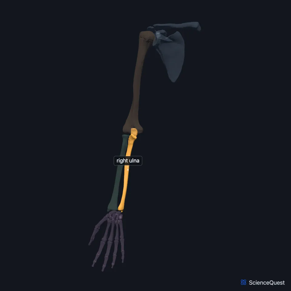
Of the two forearm bones, the ulna is the medial one. It lies parallel to the radius, and StatPearls describes it as the larger and longer of the pair. (OpenStax A&P 2e, 8.2; StatPearls, Forearm Ulna)
Open it in the Upper Limb Skeleton Explorer See it alone in 3D
Where is the ulna?
In the anatomical position, the ulna spans the forearm from the elbow to the wrist on its medial side. (StatPearls, Forearm Ulna)
Its lateral interosseous border anchors the interosseous membrane, the sheet of dense connective tissue that unites it with the radius. (OpenStax A&P 2e, 8.2; StatPearls, Forearm Ulna)
What are the parts of the ulna?
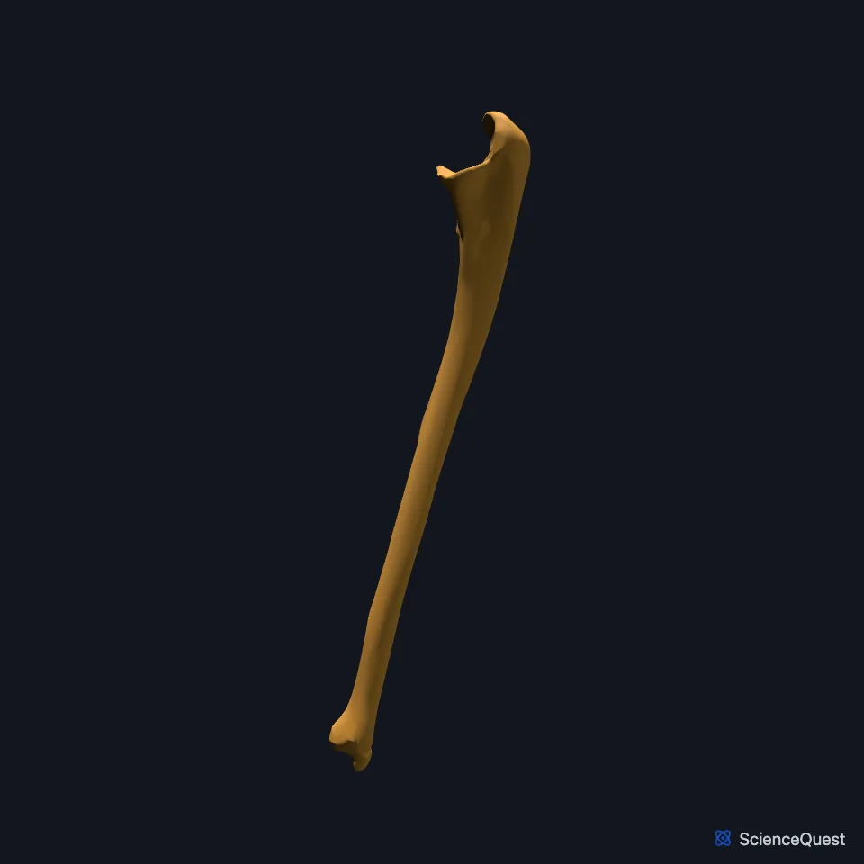
The upper end looks like a crescent wrench, with a large C-shaped trochlear notch. The coronoid process forms the lower lip of the notch, with the ulnar tuberosity below it, and the olecranon forms the curved upper and back part, its back surface lying just under the skin. (OpenStax A&P 2e, 8.2; StatPearls, Elbow Joint; Gray’s Anatomy (1918), Ulna, p. 214)
Just lateral to the trochlear notch, the small radial notch takes the head of the radius. The lower end is the small, rounded head, with a short styloid process projecting from its medial and back part. (OpenStax A&P 2e, 8.2; StatPearls, Forearm Ulna; Gray’s Anatomy (1918), Ulna, p. 214)
The ulna ossifies in three parts. The shaft starts at about eight weeks of gestation, a center appears in the head around age four, and another in the olecranon at age ten. (StatPearls, Forearm Ulna)
Which joints does the ulna form?
The trochlear notch grips the trochlea of the humerus at the humeroulnar joint, the hinge of the elbow. The radial notch and the head of the radius form the proximal radioulnar joint. (OpenStax A&P 2e, 9.6 and 8.2; StatPearls, Forearm Ulna; Gray’s Anatomy (1918), Ulna, p. 214)
At the wrist, the head of the ulna meets the ulnar notch of the radius at the distal radioulnar joint. The ulna has no joint with the carpal bones: the triquetral meets a pad of fibrocartilage instead. (OpenStax A&P 2e, 8.2; StatPearls, Forearm Ulna; Gray’s Anatomy (1918), Ulna, p. 214 and Wrist joint, p. 327)
On the medial side of the elbow, the ulnar collateral ligament runs from the medial epicondyle of the humerus to the upper ulna. It resists valgus stress, which would open the inner side of the elbow, and the lateral ligaments resist varus stress and rotation. (OpenStax A&P 2e, 9.6; StatPearls, Elbow Joint and Elbow Collateral Ligaments)
What does the ulna do?
When the elbow straightens, the olecranon fits into the olecranon fossa of the humerus. That fit takes part in flexion and extension and stops the elbow from hyperextending. (OpenStax A&P 2e, 8.2; StatPearls, Forearm Ulna)
More than a dozen muscles arise from or insert on the ulna. The brachialis inserts on the coronoid process. (StatPearls, Forearm Ulna)
| Feature | Detail and source |
|---|---|
| Bone type | Long bone (OpenStax A&P 2e, 6.2; StatPearls, Forearm Ulna; Gray’s Anatomy (1918), Classes of bones, p. 79) |
| Articulates with | Humerus (trochlea, elbow); radius (proximal and distal radioulnar joints) (OpenStax A&P 2e, 8.2; StatPearls, Forearm Ulna) |
| Carpal joints | None; the triquetral meets a fibrocartilage pad instead (OpenStax A&P 2e, 8.2; Gray’s Anatomy (1918), Ulna, p. 214 and Wrist joint, p. 327) |
Touches in this model
Measured on the BodyParts3D meshes: surfaces within 2 mm of the ulna, nearest first, 12 of 23.
- Humeral head of right extensor carpi ulnaris in contact
- Humeral head of right flexor carpi ulnaris in contact
- Humeroulnar head of right flexor digitorum superficialis in contact
- Interosseous membrane of right forearm in contact
- Lateral head of right triceps brachii in contact
- Long head of right triceps brachii in contact
- Medial head of right triceps brachii in contact
- Right abductor pollicis longus in contact
- Right anconeus in contact
- Right brachialis in contact
- Right extensor indicis in contact
- Right extensor pollicis longus in contact
Sources
- OpenStax Anatomy and Physiology 2e, section 8.2, Bones of the Upper Limb. CC BY-NC-SA 4.0. Read 2026-10-25.
- StatPearls, Anatomy, Shoulder and Upper Limb, Forearm Ulna. Mirza A. Baig; Doug W. Byerly. Last update July 24, 2023. CC BY-NC-ND 4.0. Read 2026-10-25.
- StatPearls, Anatomy, Shoulder and Upper Limb, Elbow Joint. Ryan K. Card; Jason B. Lowe; Marjorie V. Launico. Last update December 9, 2025. CC BY-NC-ND 4.0. Read 2026-10-25.
- Gray’s Anatomy of the Human Body, 20th US edition (1918), edited by Warren H. Lewis, Osteology, The Ulna (pp. 214 to 219); 1918 name: semilunar notch for trochlear notch. Henry Gray; Warren H. Lewis (editor). Public domain. Read 2026-10-25.
- OpenStax Anatomy and Physiology 2e, section 9.6, Anatomy of Selected Synovial Joints. CC BY-NC-SA 4.0. Read 2026-10-25.
- Gray’s Anatomy of the Human Body, 20th US edition (1918), edited by Warren H. Lewis, Syndesmology, Radiocarpal Articulation or Wrist-joint (pp. 327 to 328); 1918 names: navicular for scaphoid, triangular for triquetral. Henry Gray; Warren H. Lewis (editor). Public domain. Read 2026-10-25.
- StatPearls, Anatomy, Shoulder and Upper Limb, Elbow Collateral Ligaments. Michael Downing; Shane R. Sergent. Last update July 31, 2023. CC BY-NC-ND 4.0. Read 2026-10-25.
- OpenStax Anatomy and Physiology 2e, section 6.2, Bone Classification. CC BY-NC-SA 4.0. Read 2026-10-25.
- Gray’s Anatomy of the Human Body, 20th US edition (1918), edited by Warren H. Lewis, Osteology, introduction: long, short, flat and irregular bones (p. 79). Henry Gray; Warren H. Lewis (editor). Public domain. Read 2026-10-25.
The values are facts and free to use; this page’s selection and presentation are © 2026 ScienceQuest.