Radius
The radius is the lateral bone of the forearm, on the thumb side. It runs parallel to the ulna, the forearm’s other long bone. (OpenStax A&P 2e, 8.2; StatPearls, Forearm Radius)
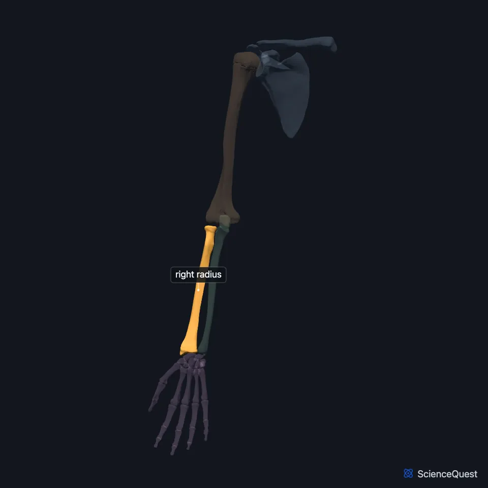
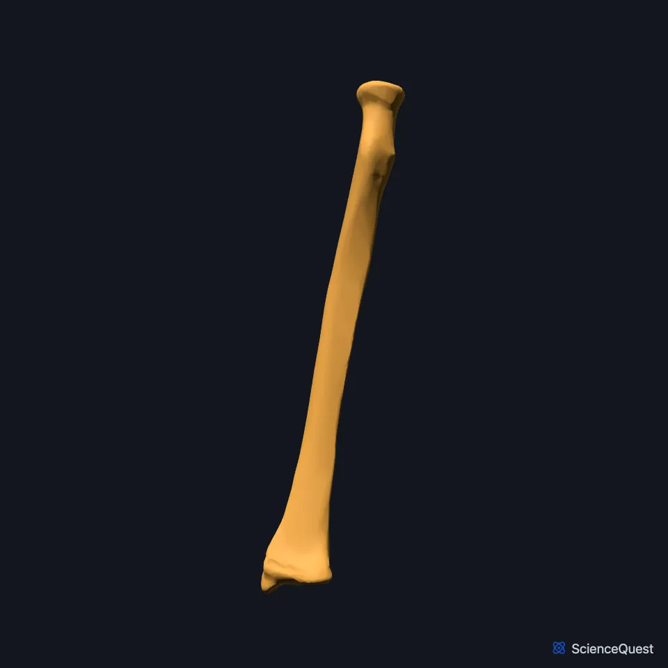
Open it in the Upper Limb Skeleton Explorer See it alone in 3D
Where is the radius?
The radius lies on the lateral side of the forearm, the part of the upper limb between the elbow and wrist joints. An interosseous membrane joins its interosseous border to the ulna. (OpenStax A&P 2e, 8.2; StatPearls, Forearm Radius; Gray’s Anatomy (1918), Radius, p. 219)
What are the parts of the radius?
The upper end is the disc-shaped head, which narrows below into the neck. Below the neck on the medial side is the oval radial tuberosity, where the biceps brachii attaches. (OpenStax A&P 2e, 8.2; StatPearls, Forearm Radius)
The shaft curves a little, convex to the lateral side. At the lower end, the ulnar notch on the medial side takes the head of the ulna, and the lateral surface runs down into the pointed styloid process. (OpenStax A&P 2e, 8.2; Gray’s Anatomy (1918), Radius, p. 219)
Which joints does the radius form?
At the elbow, the head of the radius meets the capitulum of the humerus, and the rim of the head turns in the radial notch of the ulna at the proximal radioulnar joint. (OpenStax A&P 2e, 8.2; StatPearls, Forearm Radius; Gray’s Anatomy (1918), Ulna, p. 214)
The annular ligament encircles the head of the radius and holds it against the ulna. That pivot joint lets the radius rotate in supination and pronation. (OpenStax A&P 2e, 9.6; StatPearls, Forearm Radius and Elbow Annular Ligament)
At the wrist, the ulnar notch meets the head of the ulna at the distal radioulnar joint. The lower end of the radius meets the scaphoid and the lunate at the radiocarpal, or wrist, joint. (OpenStax A&P 2e, 8.2; StatPearls, Forearm Radius; Gray’s Anatomy (1918), Wrist joint, p. 327)
What does the radius do?
The radius turns the hand. In pronation its lower end crosses over the lower end of the ulna, and reversing that movement gives supination. (StatPearls, Forearm Radius)
Muscles that attach along its shaft include the pronator teres, pronator quadratus and supinator, along with muscles of the hand that arise in the forearm. (StatPearls, Forearm Radius)
Why does the lower end of the radius break?
A fall onto an outstretched hand can break the radius just above the wrist, a Colles fracture, which is the most frequent fracture of the forearm. StatPearls puts fractures of the distal radius at 8% to 15% of all bone injuries in adults. (OpenStax A&P 2e, 8.2; StatPearls, Forearm Radius)
Does the radius vary between people?
Short, underdeveloped and absent radii have all been recorded. In proximal radioulnar synostosis, the upper ends of the radius and ulna are fused by bone, most often along their upper thirds. (StatPearls, Forearm Radius)
| Feature | Detail and source |
|---|---|
| Bone type | Long bone (OpenStax A&P 2e, 6.2; StatPearls, Forearm Radius; Gray’s Anatomy (1918), Classes of bones, p. 79) |
| Articulates with | Humerus (capitulum); ulna (proximal and distal radioulnar joints); scaphoid and lunate (wrist joint) (OpenStax A&P 2e, 8.2; StatPearls, Forearm Radius) |
| Tuberosity anchors | Biceps brachii (StatPearls, Forearm Radius) |
Touches in this model
Measured on the BodyParts3D meshes: surfaces within 2 mm of the radius, nearest first, 12 of 21.
- Humeral head of right pronator teres in contact
- Interosseous membrane of right forearm in contact
- Long head of right biceps brachii in contact
- Radial head of right flexor digitorum superficialis in contact
- Right abductor pollicis longus in contact
- Right brachialis in contact
- Right brachioradialis in contact
- Right extensor carpi radialis brevis in contact
- Right extensor carpi radialis longus in contact
- Right extensor indicis in contact
- Right extensor pollicis brevis in contact
- Right flexor digitorum profundus in contact
Sources
- OpenStax Anatomy and Physiology 2e, section 8.2, Bones of the Upper Limb. CC BY-NC-SA 4.0. Read 2026-10-25.
- StatPearls, Anatomy, Shoulder and Upper Limb, Forearm Radius. Michael M. Bair; Anoosh Zafar Gondal. Last update July 31, 2023. CC BY-NC-ND 4.0. Read 2026-10-25.
- Gray’s Anatomy of the Human Body, 20th US edition (1918), edited by Warren H. Lewis, Osteology, The Radius (pp. 219 to 221). Henry Gray; Warren H. Lewis (editor). Public domain. Read 2026-10-25.
- Gray’s Anatomy of the Human Body, 20th US edition (1918), edited by Warren H. Lewis, Osteology, The Ulna (pp. 214 to 219); 1918 name: semilunar notch for trochlear notch. Henry Gray; Warren H. Lewis (editor). Public domain. Read 2026-10-25.
- OpenStax Anatomy and Physiology 2e, section 9.6, Anatomy of Selected Synovial Joints. CC BY-NC-SA 4.0. Read 2026-10-25.
- StatPearls, Anatomy, Shoulder and Upper Limb, Elbow Annular Ligament. Brian E. Fliegel; John Ekblad; Matthew A. Varacallo. Last update July 24, 2023. CC BY-NC-ND 4.0. Read 2026-10-25.
- Gray’s Anatomy of the Human Body, 20th US edition (1918), edited by Warren H. Lewis, Syndesmology, Radiocarpal Articulation or Wrist-joint (pp. 327 to 328); 1918 names: navicular for scaphoid, triangular for triquetral. Henry Gray; Warren H. Lewis (editor). Public domain. Read 2026-10-25.
- OpenStax Anatomy and Physiology 2e, section 6.2, Bone Classification. CC BY-NC-SA 4.0. Read 2026-10-25.
- Gray’s Anatomy of the Human Body, 20th US edition (1918), edited by Warren H. Lewis, Osteology, introduction: long, short, flat and irregular bones (p. 79). Henry Gray; Warren H. Lewis (editor). Public domain. Read 2026-10-25.
The values are facts and free to use; this page’s selection and presentation are © 2026 ScienceQuest.