Scaphoid
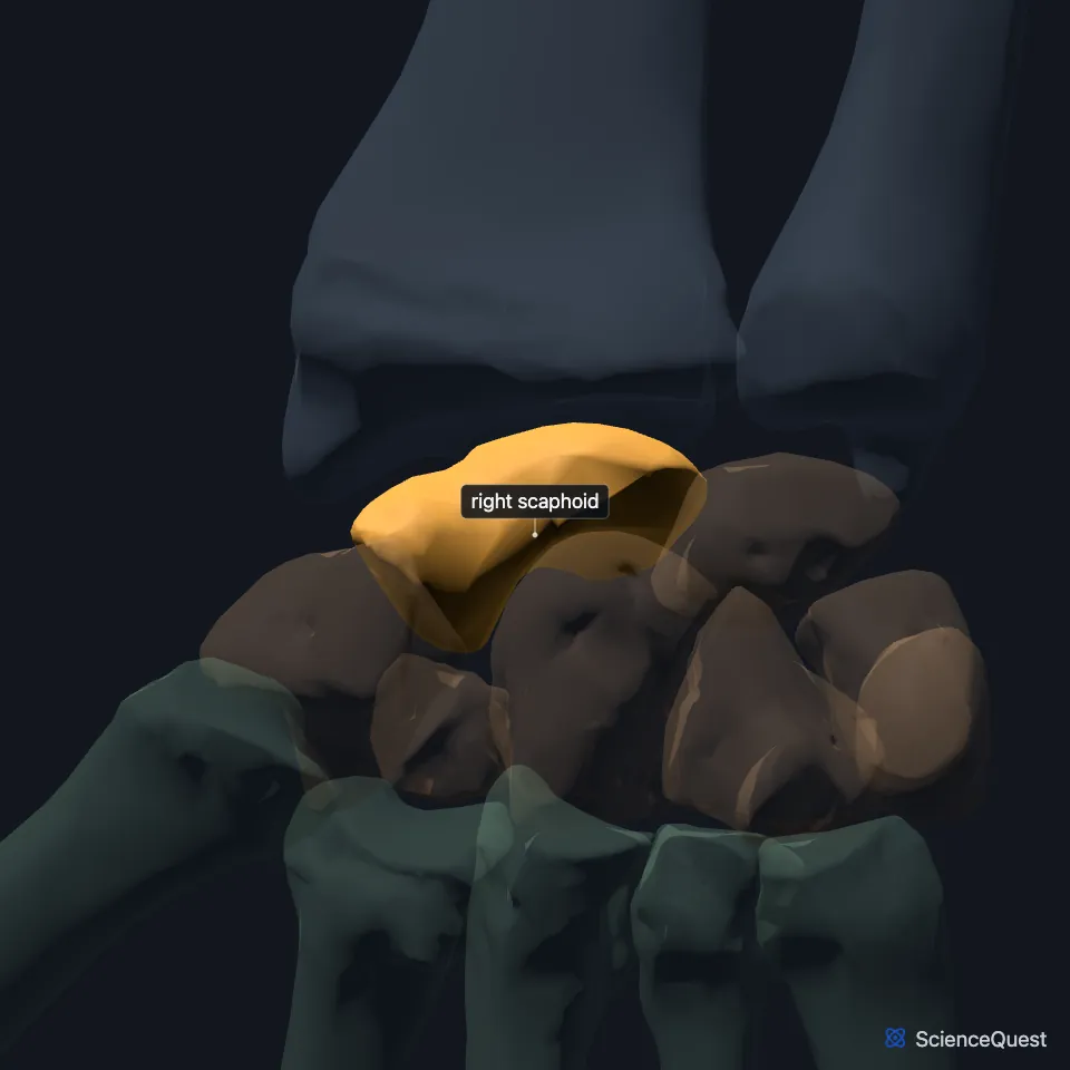
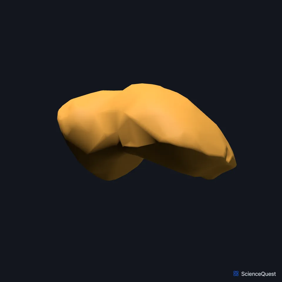
The scaphoid is a carpal bone, named for its boat shape. It forms the thumb end of the proximal row of the wrist. (OpenStax A&P 2e, 8.2; StatPearls, Hand Carpal Bones)
Open it in the Hand and Wrist Skeleton Explorer See it alone in 3D
Where is the scaphoid?
The wrist has eight carpal bones in two rows of four. The scaphoid sits at the lateral, thumb side of the proximal row, next to the lunate. (OpenStax A&P 2e, 8.2; StatPearls, Hand Carpal Bones; Gray’s Anatomy (1918), Carpus, p. 221)
In the distal row, the trapezium lies between the scaphoid and the base of the first metacarpal. (StatPearls, Hand Carpal Bones)
Which bones does the scaphoid meet?
The scaphoid and lunate meet the lower end of the radius at the radiocarpal, or wrist, joint. The triquetral joins that joint through a pad of fibrocartilage. (OpenStax A&P 2e, 8.2; Gray’s Anatomy (1918), Wrist joint, p. 327; StatPearls, Forearm Radius)
Articular cartilage covers most of its surface, which lets it bridge the joint between the two carpal rows, the midcarpal joint. (StatPearls, Hand Carpal Bones; OpenStax A&P 2e, 8.2)
The flexor retinaculum attaches laterally to the trapezium and scaphoid and medially to the hamate and pisiform. It roofs the carpal tunnel, whose walls and floor are the carpal bones. (OpenStax A&P 2e, 8.2; Gray’s Anatomy (1918), Transverse carpal ligament, p. 456 and Carpus, p. 221; StatPearls, Carpal Tunnel Syndrome and Hand Carpal Bones)
What does the scaphoid do?
The radiocarpal and midcarpal joints between them make every movement of the hand at the wrist, and the scaphoid takes part in both. (OpenStax A&P 2e, 8.2; StatPearls, Hand Carpal Bones)
The tubercles of the scaphoid and trapezium project forward. They shape the carpal tunnel and give the thumb a base from which it opposes the rest of the hand, and the thenar muscles take origin from them. (StatPearls, Hand Carpal Bones)
How does blood reach the scaphoid?
The scaphoid has a proximal and a distal pole with a waist between them. Most of its blood comes from the dorsal carpal branch of the radial artery, which enters at the dorsal ridge and flows back to supply 80% of the proximal pole. A second source, from the superficial palmar arch, enters the distal tubercle and supplies the distal pole. (StatPearls, Scaphoid Wrist Fracture)
Because the supply runs backwards, a break across the waist leaves the proximal pole at high risk of avascular necrosis. (StatPearls, Scaphoid Wrist Fracture)
Why is the scaphoid the carpal bone that breaks most?
The scaphoid is the carpal bone fractured most often, and the break often follows a fall onto the hand. StatPearls puts scaphoid fractures at 60% to 70% of carpal fractures. (OpenStax A&P 2e, 8.2; StatPearls, Scaphoid Wrist Fracture)
In a fall on an outstretched hand, its size and position make it take most of the force passed on from the radius. (StatPearls, Hand Carpal Bones)
Where the sources differ
StatPearls’ article on scaphoid fractures describes two arterial sources, one entering at the dorsal ridge and one at the distal tubercle. Its article on the carpal bones describes a single supply from the radial artery, entering at the distal part of the bone. (StatPearls, Scaphoid Wrist Fracture and Hand Carpal Bones)
StatPearls’ article on scaphoid fractures calls the scaphoid the largest of the eight carpal bones. Its article on the carpal bones names the capitate as the largest. (StatPearls, Scaphoid Wrist Fracture and Hand Carpal Bones)
| Feature | Detail and source |
|---|---|
| Bone type | Short bone, one of the eight carpals (OpenStax A&P 2e, 6.2 and 8.2; Gray’s Anatomy (1918), Classes of bones, p. 79; StatPearls, Hand Carpal Bones) |
| Position | Proximal carpal row, lateral (thumb) end (OpenStax A&P 2e, 8.2; StatPearls, Hand Carpal Bones) |
| Articulates with | Radius (radiocarpal joint); the distal carpal row (midcarpal joint) (OpenStax A&P 2e, 8.2; StatPearls, Hand Carpal Bones) |
| Blood supply | Mainly the dorsal carpal branch of the radial artery; also the superficial palmar arch (StatPearls, Scaphoid Wrist Fracture) |
Touches in this model
Measured on the BodyParts3D meshes: surfaces within 2 mm of the scaphoid, nearest first.
- Right capitate in contact
- Right lunate in contact
- Right trapezium in contact
- Right abductor pollicis brevis in contact
- Skin in contact
- Flexor retinaculum of right wrist 0.1 mm away
- Right radius 0.5 mm away
- Right trapezoid 1.4 mm away
- Right extensor indicis 1.6 mm away
Sources
- OpenStax Anatomy and Physiology 2e, section 8.2, Bones of the Upper Limb. CC BY-NC-SA 4.0. Read 2026-10-25.
- StatPearls, Anatomy, Shoulder and Upper Limb, Hand Carpal Bones. Alex Tang; Matthew A. Varacallo. Last update November 28, 2022. CC BY-NC-ND 4.0. Read 2026-10-25.
- Gray’s Anatomy of the Human Body, 20th US edition (1918), edited by Warren H. Lewis, Osteology, The Carpus (from p. 221); 1918 names: navicular for scaphoid, triangular for triquetral, greater multangular for trapezium. Henry Gray; Warren H. Lewis (editor). Public domain. Read 2026-10-25.
- Gray’s Anatomy of the Human Body, 20th US edition (1918), edited by Warren H. Lewis, Syndesmology, Radiocarpal Articulation or Wrist-joint (pp. 327 to 328); 1918 names: navicular for scaphoid, triangular for triquetral. Henry Gray; Warren H. Lewis (editor). Public domain. Read 2026-10-25.
- StatPearls, Anatomy, Shoulder and Upper Limb, Forearm Radius. Michael M. Bair; Anoosh Zafar Gondal. Last update July 31, 2023. CC BY-NC-ND 4.0. Read 2026-10-25.
- Gray’s Anatomy of the Human Body, 20th US edition (1918), edited by Warren H. Lewis, Myology, The Transverse Carpal Ligament (p. 456); 1918 names: navicular for scaphoid, greater multangular for trapezium. Henry Gray; Warren H. Lewis (editor). Public domain. Read 2026-10-25.
- StatPearls, Carpal Tunnel Syndrome. Justin O. Sevy; Reddog E. Sina; Matthew A. Varacallo. Last update October 29, 2023. CC BY-NC-ND 4.0. Read 2026-10-25.
- StatPearls, Scaphoid Wrist Fracture. Zara Hayat; Matthew A. Varacallo. Last update August 4, 2023. CC BY-NC-ND 4.0. Read 2026-10-25.
- OpenStax Anatomy and Physiology 2e, section 6.2, Bone Classification. CC BY-NC-SA 4.0. Read 2026-10-25.
- Gray’s Anatomy of the Human Body, 20th US edition (1918), edited by Warren H. Lewis, Osteology, introduction: long, short, flat and irregular bones (p. 79). Henry Gray; Warren H. Lewis (editor). Public domain. Read 2026-10-25.
The values are facts and free to use; this page’s selection and presentation are © 2026 ScienceQuest.