Occipitofrontalis
The occipitofrontalis is the broad muscle sheet of the scalp, covering one side of the vault of the skull from the occipital bone to the eyebrow. It has two bellies, the frontalis in front and the occipitalis behind, joined by a tendinous sheet, the galea aponeurotica. (Gray 1918, 4a. Muscles of the Scalp; StatPearls, Frontalis Muscle)
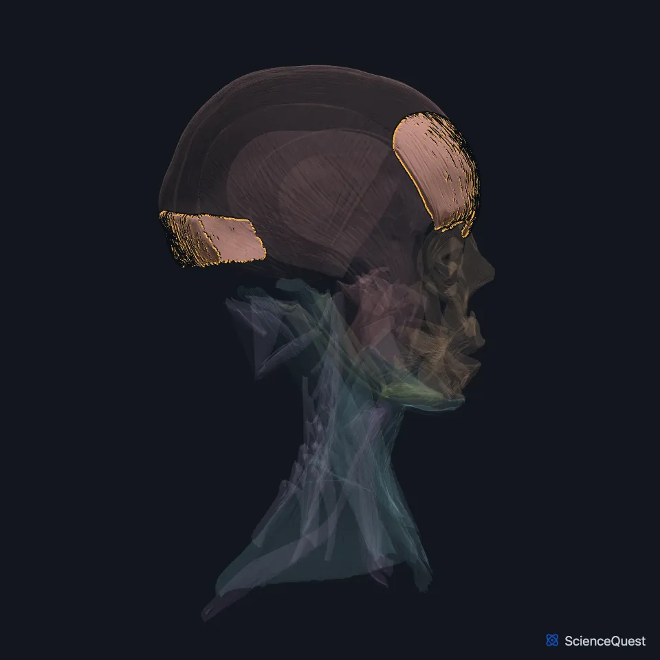
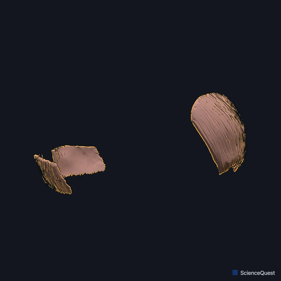
Open it in the Head and Neck Muscles Explorer See it alone in 3D
Where is the occipitofrontalis?
The occipitalis arises from the outer two thirds of the superior nuchal line of the occipital bone and from the mastoid part of the temporal bone, and it ends in the galea. (Gray 1918, 4a. Muscles of the Scalp)
The frontalis has no attachment to bone. Gray traces its fibers from the brow, where they blend with the procerus, corrugator supercilii and orbicularis oculi, up to the galea below the coronal suture. StatPearls counts the galea as its origin and the skin of the eyebrow as its insertion. (Gray 1918, 4a. Muscles of the Scalp; StatPearls, Frontalis Muscle)
The galea aponeurotica is the third of the five layers of the scalp, under the skin and the dense connective tissue and over the loose areolar tissue and the pericranium. (StatPearls, Scalp)
What is the occipitofrontalis part of?
Gray 1918 names the whole muscle the epicranius and gives occipitofrontalis as its second name. FMA uses epicranius in a wider sense: its zone of the epicranius holds the occipitofrontalis and also the temporoparietalis. (Gray 1918, 4a. Muscles of the Scalp; FMA, Occipitofrontalis and Zone of epicranius)
The frontalis forms part of the superficial musculoaponeurotic system, the fibrous layer that holds the facial muscles, which continues above the zygomatic arch. (StatPearls, Frontalis Muscle and Risorius Muscle)
What does the occipitofrontalis do?
The frontalis is the only muscle that raises the eyebrows, and it throws the skin of the forehead into horizontal lines. (StatPearls, Frontalis Muscle)
Gray describes the occipitalis holding the galea steady while the frontalis lifts the brows, which gives the face a look of surprise. Working the two bellies in turn moves the whole scalp forward and back. (Gray 1918, 4a. Muscles of the Scalp)
The procerus, corrugator supercilii and orbicularis oculi pull against the frontalis, and the balance among these muscles sets the height and shape of the eyebrow. (StatPearls, Frontalis Muscle)
What nerves and arteries supply the occipitofrontalis?
The facial nerve supplies both bellies: the frontalis through its temporal branches and the occipitalis through its posterior auricular branch. (Gray 1918, 4a. Muscles of the Scalp)
The temporal branch crosses the zygomatic arch and enters the frontalis from its deep surface. The supraorbital and supratrochlear nerves, from the ophthalmic division of the trigeminal nerve, pierce the muscle to reach the skin of the forehead. (StatPearls, Frontalis Muscle)
The supratrochlear and supraorbital arteries, from the ophthalmic artery, supply most of the frontalis from the middle. The frontal branch of the superficial temporal artery supplies its outer part. (StatPearls, Frontalis Muscle)
How much does the occipitofrontalis vary?
In up to 45% of people the right and left frontalis run into each other across the midline. In the rest a strip of galea of varying width parts them, and one dissection series found the two bellies fully separate in 15%. (StatPearls, Frontalis Muscle)
Gray notes that both bellies vary a good deal in size and in how far they attach, and that either one may be missing. (Gray 1918, 4a. Muscles of the Scalp)
Where the sources differ
Gray says the frontalis draws the scalp forward and the occipitalis draws it back. StatPearls’ frontalis article gives the reverse: the occipital belly moves the scalp forward and the frontalis moves the front of the scalp back. (Gray 1918, 4a. Muscles of the Scalp; StatPearls, Frontalis Muscle)
Gray and StatPearls’ facial muscles article give the frontalis its supply from the temporal branch of the facial nerve. StatPearls’ scalp article names the superior zygomatic nerve instead. (Gray 1918, 4a. Muscles of the Scalp; StatPearls, Facial Muscles and Scalp)
| Feature | Detail and source |
|---|---|
| Origin | Occipitalis: superior nuchal line of the occipital bone and mastoid part of the temporal bone. Frontalis: the galea aponeurotica, with no bony attachment (Gray 1918, 4a. Muscles of the Scalp; StatPearls, Frontalis Muscle) |
| Insertion | Occipitalis: the galea aponeurotica. Frontalis: the skin of the eyebrow (Gray 1918, 4a. Muscles of the Scalp; StatPearls, Frontalis Muscle) |
| Action | Frontalis raises the eyebrows and lines the forehead; the two bellies move the scalp (StatPearls, Frontalis Muscle; Gray 1918, 4a. Muscles of the Scalp) |
| Nerve | Facial nerve (CN VII): temporal branches to the frontalis, posterior auricular branch to the occipitalis (Gray 1918, 4a. Muscles of the Scalp) |
Touches in this model
Measured on the BodyParts3D meshes: surfaces within 2 mm of the occipitofrontalis, nearest first, 12 of 21.
- Aponeurosis of epicranius in contact
- Left corrugator supercilii in contact
- Left procerus in contact
- Left temporalis in contact
- Left temporoparietalis in contact
- Palpebral part of left orbicularis oculi in contact
- Palpebral part of right orbicularis oculi in contact
- Right corrugator supercilii in contact
- Right procerus in contact
- Right temporalis in contact
- Right temporoparietalis in contact
- Frontal bone in contact
Sources
Sources read between and
- Gray’s Anatomy of the Human Body, 20th US edition (1918), IV. Myology, 4. The Fasciæ and Muscles of the Head, a. The Muscles of the Scalp (1918 terms: Epicranius for occipitofrontalis). Henry Gray. Public domain. Read .
- StatPearls, Anatomy, Head and Neck; Frontalis Muscle. Kenneth Pessino; Jay Patel; Bhupendra C. Patel. Last update . CC BY-NC-ND 4.0. Read .
- StatPearls, Anatomy, Head and Neck, Scalp. Jahan Tajran; Amanda A. Gosman. Last update . CC BY-NC-ND 4.0. Read .
- Foundational Model of Anatomy, FMA9624, Occipitofrontalis (read on EBI OLS). FMA release terms, University of Washington. Read .
- Foundational Model of Anatomy, FMA46752, Zone of epicranius (read on EBI OLS). FMA release terms, University of Washington. Read .
- StatPearls, Anatomy, Head and Neck, Risorius Muscle. Alex M. Germann; Yasir Al Khalili. Last update . CC BY-NC-ND 4.0. Read .
- StatPearls, Anatomy, Head and Neck: Facial Muscles. Adegbenro O. Fakoya; Marc H. Hohman; Katherine E. Westbrook; Matthew A. Varacallo. Last update . CC BY-NC-ND 4.0. Read .
The values are facts and free to use; this page’s selection and presentation are © 2026 ScienceQuest.