Rectus capitis posterior minor
The rectus capitis posterior minor is one of the four suboccipital muscles, at the back of the neck just below the occipital bone. It runs from the back of the atlas up to the skull. (StatPearls, Suboccipital Muscles; Gray 1918, 6b. Suboccipital Muscles)
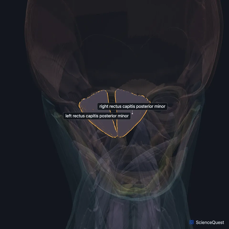
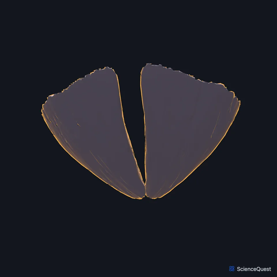
Open it in the Head and Neck Muscles Explorer See it alone in 3D
Where is the rectus capitis posterior minor?
As the two rectus capitis posterior major muscles rise and spread apart, they leave a triangular space between them. The minor muscles of the two sides lie in that space. (Gray 1918, 6b. Suboccipital Muscles)
It is not one of the borders of the suboccipital triangle. The major muscle and the two oblique muscles form those borders, and the triangle holds the suboccipital nerve, part of the vertebral artery and a venous plexus. (Gray 1918, 6b. Suboccipital Muscles; StatPearls, Suboccipital Muscles)
Where does the rectus capitis posterior minor attach?
It arises by a narrow, pointed tendon from the tubercle on the posterior arch of the atlas. It widens as it rises and inserts on the medial part of the inferior nuchal line of the occipital bone and the bone between that line and the foramen magnum. (Gray 1918, 6b. Suboccipital Muscles; StatPearls, Suboccipital Muscles)
That posterior tubercle is the small remnant of a spinous process. Gray’s Anatomy (1918) notes that its small size keeps it from getting in the way of movement between the atlas and the skull. (Gray 1918, 3a. 1. The Cervical Vertebrae)
What does the rectus capitis posterior minor do?
The suboccipital muscles hold the head and neck up and allow extension and turning. Gray’s Anatomy (1918) says the two recti draw the head backward. (StatPearls, Suboccipital Muscles; Gray 1918, 6b. Suboccipital Muscles)
Like the other three suboccipital muscles, it is supplied by the suboccipital nerve, the dorsal ramus of the first cervical spinal nerve. (StatPearls, Suboccipital Muscles)
What is the rectus capitis posterior minor linked to?
The vertebral artery runs deep to the suboccipital muscles, in a groove on the posterior arch of the atlas, and gives off branches that supply them. StatPearls names that closeness, and surgical access to the posterior cranial fossa, as the reasons these muscles matter in the clinic. (Gray 1918, 6b. Suboccipital Muscles; StatPearls, Suboccipital Muscles)
In a 1995 study of 11 cadaver specimens, Hack and colleagues found a bridge of connective tissue joining this muscle to the spinal dura mater where the atlas meets the skull. It was there in every specimen. StatPearls reports the hypothesis that injury to this muscle matters most in lasting pain after whiplash, and that a tear of it after trauma could cause cervicogenic headache. (Hack et al. 1995, Spine; StatPearls, Suboccipital Muscles)
Is the rectus capitis posterior minor ever missing?
Rarely. StatPearls cites a case with no minor muscle on one side and two rectus capitis posterior major muscles in its place, and another with a third rectus capitis muscle in the region. (StatPearls, Suboccipital Muscles)
Where the sources differ
The sources differ on rotation. StatPearls lists the muscle’s action as extending the neck and turning the head to the same side, while Gray’s Anatomy (1918) has both recti draw the head backward and gives the turning action to the rectus capitis posterior major. (StatPearls, Suboccipital Muscles; Gray 1918, 6b. Suboccipital Muscles)
| Feature | Detail and source |
|---|---|
| Origin | Tubercle on the posterior arch of the atlas (Gray 1918, 6b. Suboccipital Muscles; StatPearls, Suboccipital Muscles) |
| Insertion | Medial part of the inferior nuchal line of the occipital bone, and the bone below it (Gray 1918, 6b. Suboccipital Muscles) |
| Action | Draws the head backward (Gray 1918, 6b. Suboccipital Muscles; StatPearls, Suboccipital Muscles) |
| Nerve | Suboccipital nerve, the dorsal ramus of C1 (StatPearls, Suboccipital Muscles) |
Touches in this model
Measured on the BodyParts3D meshes: surfaces within 2 mm of the rectus capitis posterior minor, nearest first.
- Left rectus capitis posterior major in contact
- Right rectus capitis posterior major in contact
- Atlas in contact
- Descending part of left trapezius in contact
- Descending part of right trapezius in contact
- Occipital bone in contact
- Set of interspinales cervicis in contact
- Skin in contact
- Left semispinalis capitis 0.3 mm away
- Right semispinalis capitis 0.3 mm away
- Left cervical rotator 1.6 mm away
- Right cervical rotator 1.6 mm away
Sources
Sources read between and
- StatPearls, Anatomy, Head and Neck, Suboccipital Muscles. Tom George; Prasanna Tadi. Last update . CC BY-NC-ND 4.0. Read .
- Gray’s Anatomy of the Human Body, 20th US edition (1918), IV. Myology, 6b. The Suboccipital Muscles, actions of the deep back muscles (1918 terms: Sacrospinalis for erector spinae; odontoid process for the dens; posterior occipito-atlantal membrane for the posterior atlanto-occipital membrane; Rectus capitis posticus major and minor given as second names). Henry Gray. Public domain. Read .
- Gray’s Anatomy of the Human Body, 20th US edition (1918), II. Osteology, 3a. 1. The Cervical Vertebrae. Henry Gray. Public domain. Read .
- Spine, Anatomic relation between the rectus capitis posterior minor muscle and the dura mater, Spine 20, pages 2484 to 2486 (abstract, PMID 8610241). Hack GD, Koritzer RT, Robinson WL, Hallgren RC, Greenman PE. Abstract read through NCBI E-utilities; no reuse licence stated. Read .
The values are facts and free to use; this page’s selection and presentation are © 2026 ScienceQuest.