Rectus capitis posterior major
The rectus capitis posterior major is one of the four suboccipital muscles, which lie at the back of the neck just below the occipital bone. (StatPearls, Suboccipital Muscles)
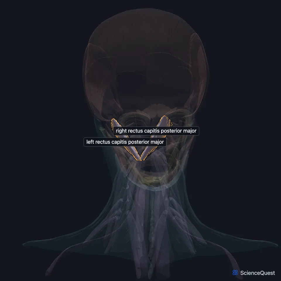
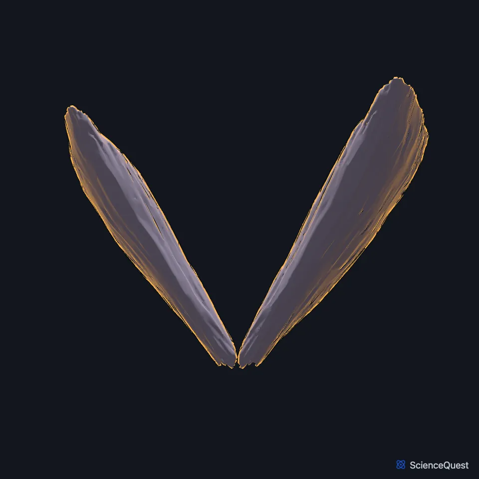
Open it in the Head and Neck Muscles Explorer See it alone in 3D
Where is the rectus capitis posterior major?
It arises by a pointed tendon from the spinous process of the axis and widens as it rises to the lateral part of the inferior nuchal line of the occipital bone and the bone just below it. (Gray 1918, 6b. Suboccipital Muscles; StatPearls, Suboccipital Muscles)
The left and right muscles spread apart as they rise and leave a triangular gap between them, where the rectus capitis posterior minor lies. (Gray 1918, 6b. Suboccipital Muscles)
What does the rectus capitis posterior major do?
It draws the head backward. Because it runs at a slant, it also turns the skull and atlas around the dens of the axis, so the face turns to the same side. (Gray 1918, 6b. Suboccipital Muscles; StatPearls, Suboccipital Muscles)
The suboccipital muscles also work as postural muscles that steady the head on the neck. (StatPearls, Suboccipital Muscles)
What does the rectus capitis posterior major border?
It forms the upper medial side of the suboccipital triangle, with the obliquus capitis superior and obliquus capitis inferior on the other two sides. The triangle holds the third segment of the vertebral artery and the suboccipital nerve. (Gray 1918, 6b. Suboccipital Muscles; StatPearls, Suboccipital Muscles)
The suboccipital nerve, the dorsal ramus of the first cervical nerve, supplies it, and a muscular branch from the third segment of the vertebral artery brings its blood. The semispinalis capitis covers the group from behind. (StatPearls, Suboccipital Muscles)
Are the suboccipital muscles linked to headaches?
StatPearls notes connections between the suboccipital muscles and the dura mater, called the myodural bridge. Researchers have proposed that tension in these muscles pulls on the dura and helps cause cervicogenic headache. (StatPearls, Suboccipital Muscles)
How does the rectus capitis posterior major vary?
One case report describes a person with no rectus capitis posterior minor on one side and two rectus capitis posterior major muscles in its place. (StatPearls, Suboccipital Muscles)
| Feature | Detail and source |
|---|---|
| Origin | Spinous process of the axis (C2) (Gray 1918, 6b. Suboccipital Muscles; StatPearls, Suboccipital Muscles) |
| Insertion | Lateral part of the inferior nuchal line of the occipital bone, and the bone just below it (Gray 1918, 6b. Suboccipital Muscles) |
| Action | Extends the head and turns the face to the same side (StatPearls, Suboccipital Muscles; Gray 1918, 6b. Suboccipital Muscles) |
| Nerve | Suboccipital nerve (dorsal ramus of C1) (StatPearls, Suboccipital Muscles) |
Touches in this model
Measured on the BodyParts3D meshes: surfaces within 2 mm of the rectus capitis posterior major, nearest first, 12 of 16.
- Left obliquus capitis inferior in contact
- Left obliquus capitis superior in contact
- Left rectus capitis posterior minor in contact
- Right obliquus capitis inferior in contact
- Right obliquus capitis superior in contact
- Right rectus capitis posterior minor in contact
- Axis in contact
- Descending part of left trapezius in contact
- Descending part of right trapezius in contact
- Left semispinalis capitis in contact
- Occipital bone in contact
- Right semispinalis capitis in contact
Sources
Sources read
- StatPearls, Anatomy, Head and Neck, Suboccipital Muscles. Tom George; Prasanna Tadi. Last update . CC BY-NC-ND 4.0. Read .
- Gray’s Anatomy of the Human Body, 20th US edition (1918), IV. Myology, 6b. The Suboccipital Muscles, actions of the deep back muscles (1918 terms: Sacrospinalis for erector spinae; odontoid process for the dens; posterior occipito-atlantal membrane for the posterior atlanto-occipital membrane; Rectus capitis posticus major and minor given as second names). Henry Gray. Public domain. Read .
The values are facts and free to use; this page’s selection and presentation are © 2026 ScienceQuest.