Mitosis and Meiosis Explorer
Mitosis makes two identical diploid cells; meiosis makes four different haploid ones. Step through each stage and count chromosomes for 2n up to 46.
Simulator
Left and right arrow keys step between stages, and Home and End jump to the first and last. Space plays and pauses.
Stage 1 of 9, Interphase, G1. The cell grows. Each chromosome is one long DNA molecule, spread out as chromatin inside the nucleus.
- Chromosomes per cell 2n: one set from each parent.
- 4
- Chromatids per cell One DNA molecule per chromosome. Some books count no chromatids until replication, keeping the word for half of a copied chromosome.
- 4
- DNA per cell In C, the DNA in one unreplicated set of chromosomes, so a diploid cell starts at 2C.
- 2C
- Cells Still one cell.
- 1
- DNA per cell (C)
- Chromosomes per cell (n)
| Step | Stage | Cells | Chromosomes | Chromatids | DNA |
|---|---|---|---|---|---|
| 1 | 1 | 4 | 4 | 2C | |
| 2 | 1 | 4 | 4 to 8 | 2C to 4C | |
| 3 | 1 | 4 | 8 | 4C | |
| 4 | 1 | 4 | 8 | 4C | |
| 5 | 1 | 4 | 8 | 4C | |
| 6 | 1 | 4 | 8 | 4C | |
| 7 | 1 | 8 | 8 | 4C | |
| 8 | 1 | 8 | 8 | 4C | |
| 9 | 2 | 4 | 4 | 2C |
| Feature | Mitosis | Meiosis |
|---|---|---|
| Divisions | 1 | 2 |
| Cells made | 2 | 4 |
| Chromosomes in each | 4 | 2 |
| Chromatids in each | 4 | 2 |
| DNA in each | 2C | 1C |
| Homologous pairs in each | 2 | None |
| Identical to the parent | Yes | No |
| Homologues pair and cross over | No | Yes, in prophase I |
| What separates at anaphase | Sister chromatids | Homologues, then sister chromatids |
| Kinds of cell from assortment | 1 | 4 |
Citing this tool
Last updated . Add the date you accessed it as well, which a citation of a page that can change asks for. If a specific result matters, cite the permalink from the tool’s share row instead of this page: it reproduces the exact parameters.
The equation
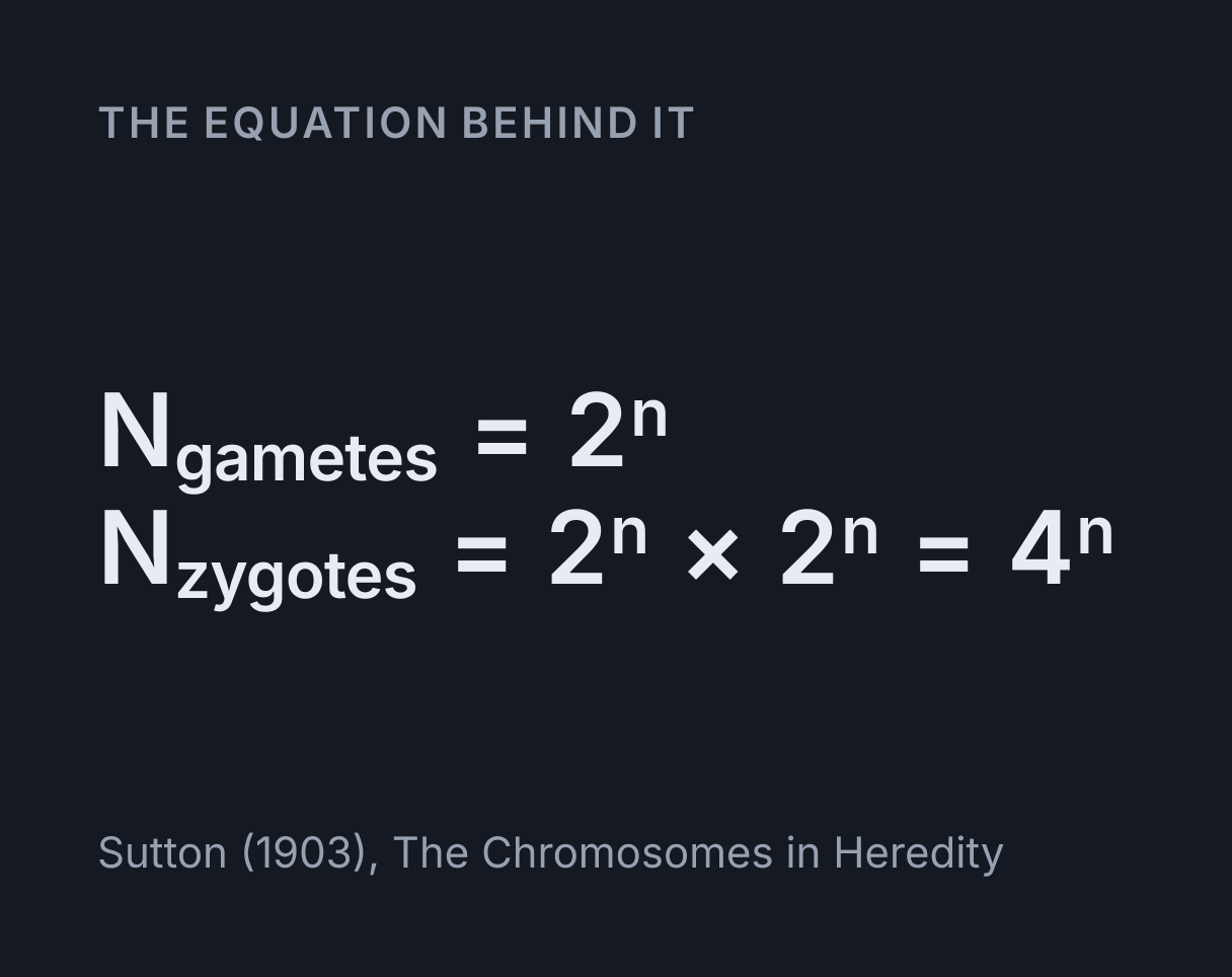
Sutton (1903), The Chromosomes in Heredity
Mitosis makes two identical cells; meiosis makes four different ones
Mitosis divides one cell into two genetically identical daughter cells, each with the
parent’s chromosome number, the full diploid number 2n in a body cell. Meiosis divides a
diploid cell twice to make four genetically different cells, each with the haploid number,
n: in a human, 46 chromosomes in each
daughter of mitosis and 23 in each product of meiosis. The pairs line up independently at
metaphase I, so one person can make 2^n kinds of gamete before crossing over is
counted.
Every count on this page follows two rules: chromosomes = centromeres and
chromatids = DNA molecules, each chromatid being one
DNA double helix. Copying the DNA in S phase
doubles the chromatids and leaves the chromosome number alone. Anaphase doubles the
chromosome number, because once the sisters separate each chromatid counts as a chromosome
in its own right. Cytokinesis then shares everything between two cells.
How to use it
Choose mitosis or meiosis and a diploid number, then press Play to watch the division stage by stage, or step with Previous and Next, the arrow keys or the rows of the stage table. The readouts give what each cell holds at the stage on screen, and the table lists every stage at once for the 2n you chose, so a question such as how many chromatids a cell has at metaphase II can be read straight off it. The graph plots the DNA per cell through the whole division, with the chromosome number drawn against it.
In meiosis the colours carry the genetics: red chromosomes came from the mother and blue from the father. Shuffle picks another way for the pairs to line up at metaphase I, which changes the four cells made at the end, and crossing over leaves some chromatids with a tip of the other colour. A population of cells dividing by mitosis grows at the rate the cell doubling time calculator works out.
Worked example: a human cell, 2n = 46
| Stage | Chromosomes | Chromatids | DNA |
|---|---|---|---|
| G1, before replication | 46 | 46 | 2C |
| G2 to metaphase | 46 | 92 | 4C |
| Anaphase and telophase of mitosis | 92 | 92 | 4C |
| Each daughter of mitosis | 46 | 46 | 2C |
| Prophase I to telophase I | 46 | 92 | 4C |
| Each cell after meiosis I | 23 | 46 | 2C |
| Anaphase II | 46 | 46 | 2C |
| Each cell after meiosis II | 23 | 23 | 1C |
Two rows catch most people out. A cell that has finished meiosis I has 23 chromosomes but 46 chromatids, and it is haploid even though it holds as much DNA as a body cell in G1. In anaphase II it has 46 chromosomes again, but they are the separated halves of 23, with no homologous pairs among them.
The same 23 pairs set the variety. Each pair can face either pole at metaphase I, so one
person can make 2^23 = 8,388,608 kinds of gamete before crossing over is
counted, and two parents can combine them in 4^23 ways, about 7.04 × 10¹³. Set
2n to 46 and the tool reproduces every number in the table.
Where the variety comes from
Independent assortment. At metaphase I each homologous pair lines up with one member facing
each pole, and which way round it lands is independent of every other pair. Walter Sutton
pointed out in 1903 that this alone lets one individual make 2^n kinds of
gamete, and tabulated the numbers from 2 to 36 chromosomes. He illustrated them with the
lowest estimate of the human count then published, 16; the count of 46 was established by
Tjio and Levan in 1956.
The tool counts arrangements the careful way. With n pairs there are 2^n ways to
orient them, but an arrangement and its mirror image send the same two sets of chromosomes to
the two poles, so there are 2^(n − 1) distinct arrangements, each making two
kinds of cell. For a human cell that is 4,194,304 arrangements and 8,388,608 kinds of gamete.
Mendel’s ratios follow from the same two facts, and the Punnett square calculator works them out: each gamete gets one chromosome of every pair, so one allele of each gene, and genes on different chromosomes assort independently because their pairs do.
Crossing over. In prophase I a maternal and a paternal chromatid of each pair break at the same point and rejoin the other way round, so a chromosome can leave carrying genes from both parents. In most species every pair needs at least one crossover, because the chiasma it forms is what holds the homologues together until anaphase I. Watch the colours: each side of a bivalent stays one colour until the homologues are pulled apart, because cohesin still holds each exchanged tip beside its old sister, and only then do the recombinant chromatids show a tip of the other colour.
The stages in order
Interphase comes first: G1, then S, when the DNA is copied, then G2. Mitosis itself runs prophase, prometaphase, metaphase, anaphase and telophase, and cytokinesis then divides the cell. Some courses list only four stages and count prometaphase as part of prophase. The stages, the counting rules and the captions here follow OpenStax Biology 2e, sections 10.2 and 11.1.
- Prophase: chromosomes condense and the spindle starts to form.
- Prometaphase: the nuclear envelope breaks down and kinetochores attach to the spindle.
- Metaphase: chromosomes line up on the metaphase plate.
- Anaphase: sister chromatids separate and move to opposite poles.
- Telophase: nuclear envelopes reform around the two sets and the chromosomes decondense.
Meiosis passes through the same stages twice, as meiosis I and meiosis II, with no S phase in between, and the tool folds prometaphase into the stages either side of it. Prophase I is long and is usually split into leptotene, zygotene, pachytene, diplotene and diakinesis. The three prophase I steps here are pairing, which covers leptotene and zygotene, crossing over, which is pachytene, and chiasmata, which covers diplotene and diakinesis.
What the drawing leaves out
- Real timing. Each stage gets a few seconds so it can be watched, and none of the durations are to scale. A human egg, for instance, stops in prophase I before birth and can stay there for decades.
- Real sizes. Chromosomes are drawn to fit the cell, each pair a different length with its centromere in a different place so that the pairs can be told apart. They are not to scale for any species. When there are too many to fit end to end across the cell, the metaphase plate is drawn as a band several chromosomes deep, standing for a plate seen edge on.
- Variable crossovers. Every pair crosses over exactly once, on its long arm, between its two inner chromatids. Real pairs can have more than one crossover, at varying places.
- Egg formation. The four equal cells at the end match sperm formation, which happens in the testes the male reproductive organs explorer shows in 3D. In egg formation the divisions are unequal, giving one egg and small polar bodies, and meiosis II is completed only if the egg is fertilised.
- Plant meiosis. Meiosis is drawn in an animal cell. In a plant it makes haploid spores rather than gametes, and the gametes come later from the gametophytes those spores grow into.
- Mistakes. Nondisjunction, when homologues or sister chromatids fail to separate, leaves cells with an extra or a missing chromosome, and is not modelled.
Common mistakes
- Counting chromatids as chromosomes. A replicated chromosome is one chromosome with two chromatids. S phase doubles the DNA, not the chromosome number.
- Halving the chromosome number in meiosis II. It halves in meiosis I, when the homologues separate. Meiosis II halves the DNA in each cell and leaves the chromosome number at n.
- Calling the cells after meiosis I diploid. They hold 2C of DNA, but only one chromosome of each pair, so they are haploid.
- Putting DNA replication in prophase. It happens in S phase, before division starts, and not at all between meiosis I and meiosis II.
- Treating homologous chromosomes as copies. Homologues carry the same genes in the same places, one from each parent, in versions that can differ. Sister chromatids are the copies.
- Crossing over between sisters. The exchange that makes new combinations is between non-sister chromatids, one maternal and one paternal.
Model and assumptions
- Method
- Exact expression, no time stepping
- Repeatability
- Deterministic. The same link gives the same numbers on any machine.
What it assumes
- Every chromatid is tracked through both divisions, so the counts at each stage and the four cells at the end are computed from the arrangement and the crossovers rather than drawn by hand.
- Chromosomes are counted by centromere and chromatids by DNA molecule, per cell, and a dividing cell counts as one until its furrow closes.
- Which way each homologous pair faces at metaphase I is set by the arrangement in the link, and with crossing over on each pair crosses over exactly once, on its long arm, between its two inner chromatids.
- Meiosis is drawn with equal divisions in an animal cell, as in sperm formation, and every stage is given a few seconds rather than its real duration.
Where it stops holding. Real crossovers vary in number and position, egg formation divides unequally, and errors such as nondisjunction, which leave a cell with an extra or a missing chromosome, are not modelled.
Numerical accuracy
No method error to report: the result is a closed-form expression evaluated directly, with no time stepping to accumulate error. What remains is double-precision rounding, of order one part in 10^16 per operation.
Common questions
What is the difference between mitosis and meiosis?
Mitosis makes two cells identical to the parent, each with the same number of chromosomes, the full diploid set in a body cell, while meiosis makes four cells with half that number, each genetically different. Mitosis is one division and is how the body grows and replaces cells. Meiosis is two divisions in a row with no DNA copying between them, and in animals it makes sperm and eggs. The differences come from meiosis I: homologous chromosomes pair up, swap pieces by crossing over and are then pulled apart, whereas mitosis only ever separates sister chromatids.
What are the stages of mitosis?
Prophase, prometaphase, metaphase, anaphase and telophase, followed by cytokinesis, which divides the cell in two. The chromosomes condense in prophase, attach to the spindle in prometaphase, line up in metaphase, separate into sister chromatids in anaphase and decondense inside two new nuclei in telophase. Some courses list only four stages and count prometaphase as part of prophase. Before any of it the cell passes through interphase: G1, S phase, when the DNA is copied, and G2.
How do you count chromosomes and chromatids?
Count centromeres for chromosomes and DNA molecules for chromatids. A replicated chromosome is still one chromosome, with two sister chromatids joined at the centromere, so copying the DNA in S phase doubles the chromatids but not the chromosomes. The chromosome number only doubles at anaphase, when the sisters separate and each counts as a chromosome in its own right. So a human cell in G2 has 46 chromosomes and 92 chromatids, in anaphase of mitosis it briefly has 92 chromosomes, and after meiosis I each cell has 23 chromosomes but still 46 chromatids. Some books keep the word chromatid for half of a copied chromosome, and so count none before S phase.
How many different gametes can one person make?
At least 2 to the power 23, which is 8,388,608, from independent assortment alone. Each of the 23 pairs lines up at metaphase I with either member facing either pole, independently of the others, so the choices multiply. Two parents can combine those gametes in 8,388,608 squared ways, about 70 trillion. Crossing over in prophase I adds far more variety on top.
What is the difference between crossing over and independent assortment?
Crossing over mixes genes within a chromosome, and independent assortment mixes whole chromosomes. Crossing over happens in prophase I, when a maternal and a paternal chromatid of a pair break at the same point and rejoin the other way round, so a chromosome can leave carrying genes from both parents. Independent assortment happens at metaphase I, when each pair’s orientation on the plate is set by chance, so which parent’s copy of each chromosome a gamete receives is independent from one pair to the next.