Protein Synthesis Simulator
Protein synthesis is transcription, DNA copied into mRNA, then translation, where a ribosome adds one amino acid per codon. Step through your own gene.
Simulator
Right and left arrow keys go forward and back a step, Home and End to the start and the end. Space plays and pauses.
The gene, in the nucleus, as a double helix: the coding strand on top, read 5′ to 3′ from left to right, and the template strand below it, 96 base pairs. Press Play, or Next to go one step at a time.
Step 0 of 197Codons, anticodons and amino acids
Protein, 31
amino acids Met-Gly-Ile-Asn-Thr-Arg-Glu-Leu-Phe-Leu-Asn-Phe-Thr-Ile-Val-Leu-Ile-Thr-Val-Ile-Leu-Met-Trp-Leu-Leu-Val-Arg-Ser-Tyr-Gln-Tyr
Each codon of the reading frame, the anticodon that pairs with it and the amino acid it codes for. Anticodons are lined up under their codons, so they read 3′ to 5′; the 5′ to 3′ form, which tRNAs are named by, is in brackets. Each is the exact complement: a cell may read a codon with a tRNA that pairs more loosely at the third base, which is called wobble. Choose a number to go to the step where that codon is read.
| # | Codon, 5′ to 3′ | Anticodon, 3′ to 5′ | Amino acid |
|---|---|---|---|
AUG | UAC (CAU) | Met, methionine | |
GGG | CCC (CCC) | Gly, glycine | |
AUA | UAU (UAU) | Ile, isoleucine | |
AAC | UUG (GUU) | Asn, asparagine | |
ACC | UGG (GGU) | Thr, threonine | |
CGG | GCC (CCG) | Arg, arginine | |
GAG | CUC (CUC) | Glu, glutamic acid | |
CUG | GAC (CAG) | Leu, leucine | |
UUU | AAA (AAA) | Phe, phenylalanine | |
CUC | GAG (GAG) | Leu, leucine | |
AAC | UUG (GUU) | Asn, asparagine | |
UUC | AAG (GAA) | Phe, phenylalanine | |
ACU | UGA (AGU) | Thr, threonine | |
AUU | UAA (AAU) | Ile, isoleucine | |
GUC | CAG (GAC) | Val, valine | |
UUG | AAC (CAA) | Leu, leucine | |
AUU | UAA (AAU) | Ile, isoleucine | |
ACG | UGC (CGU) | Thr, threonine | |
GUU | CAA (AAC) | Val, valine | |
AUU | UAA (AAU) | Ile, isoleucine | |
CUU | GAA (AAG) | Leu, leucine | |
AUG | UAC (CAU) | Met, methionine | |
UGG | ACC (CCA) | Trp, tryptophan | |
CUC | GAG (GAG) | Leu, leucine | |
CUU | GAA (AAG) | Leu, leucine | |
GUG | CAC (CAC) | Val, valine | |
AGG | UCC (CCU) | Arg, arginine | |
UCC | AGG (GGA) | Ser, serine | |
UAU | AUA (AUA) | Tyr, tyrosine | |
CAG | GUC (CUG) | Gln, glutamine | |
UAC | AUG (GUA) | Tyr, tyrosine | |
UGA | none | stop, read by eRF1 |
- Nonpolar, aliphatic
- Aromatic
- Polar, uncharged
- Positively charged
- Negatively charged
All six reading frames of this sequence, with its open reading frames marked: open it in the DNA to Protein Translator.
- Protein From the AUG at base 1 to the stop codon UGA at bases 94 to 96, which adds no amino acid.
- 31 amino acids
- Chain so far Amino acids joined so far, the first one included.
- 0 amino acids
- Codon being read Nothing is being decoded at this step.
- none
- Its anticodon No tRNA is pairing with a codon at this step.
- none
- mRNA As typed, before the cap and the tail are added.
- 96 bases
- Transcription in a cell The bases typed divided by the 50 to 100 nucleotides a second that RNA polymerase II manages in mammalian cells (Milo and Phillips 2015). Elongation only.
- 0.96 to 1.9 s
- Translation in a cell The amino acids divided by the 6 a second a ribosome adds in mammalian cells (Milo and Phillips 2015). Elongation only.
- 5.2 s
Citing this tool
Last updated . Add the date you accessed it as well, which a citation of a page that can change asks for. If a specific result matters, cite the permalink from the tool’s share row instead of this page: it reproduces the exact parameters.
The equation
Elongation rates from Milo and Phillips (2015), Cell Biology by the Numbers
What protein synthesis is
Protein synthesis is how a cell makes a protein from a gene, in two stages. In
transcription, RNA polymerase copies the gene’s template strand into
messenger RNA; in translation, a ribosome reads that mRNA three bases at a
time, from the start codon AUG to a stop codon, and joins one amino acid for
every codon before the stop.
Three rules do all the work, and the tool applies them to whatever you type:
-
mRNA = coding strand with U in place of T, because each mRNA base pairs with the template base opposite it: A with U, T with A, G with C and C with G. -
amino acids = (bases from the A of AUG to the last base of the stop codon) ÷ 3 − 1, because the stop codon adds none. -
anticodon = the complement of the codon, read the other way, because tRNA and mRNA pair antiparallel: under 5′-AUG-3′ sits 3′-UAC-5′.
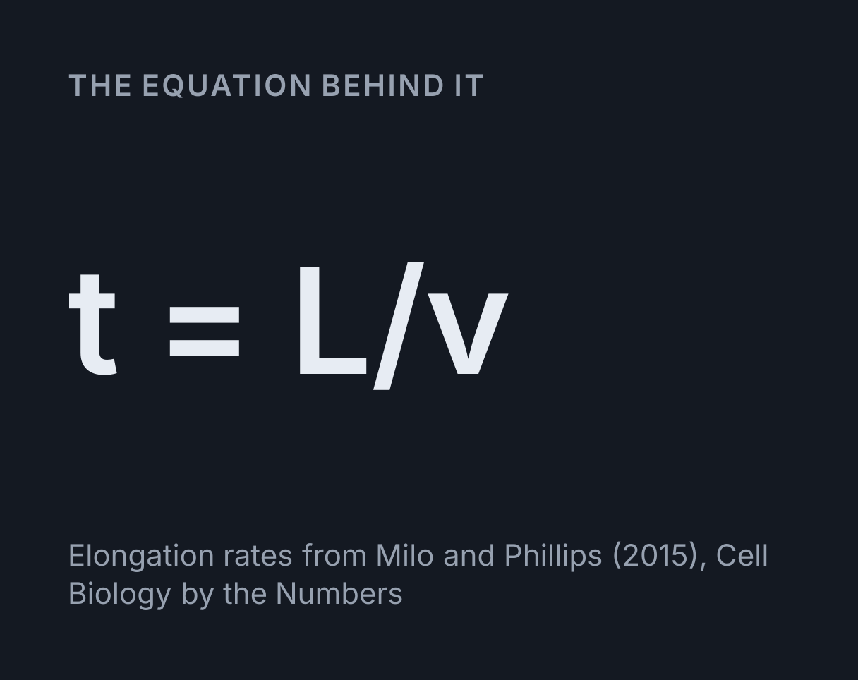
The equation above, t = L ÷ v, is the one thing timed: the length made divided
by the elongation rate, which gives the readouts that say how long a real cell would take.
How to use it
Type or paste a DNA or mRNA sequence, or choose an example, then say which strand it is. The coding strand reads like the mRNA; the template strand is the one RNA polymerase reads, and it can be typed written either way round, 3′ to 5′ or 5′ to 3′. Choose a eukaryotic cell to see the mRNA capped, tailed and exported from the nucleus, or a bacterium, which has no nucleus.
Press Play to watch the whole process, or Next to go one event at a time: each base the polymerase adds, each tRNA that arrives, each peptide bond and each move of the ribosome. The caption under the scene says what the current step does, and the codon table lists every codon, its anticodon and its amino acid; choose a codon’s number to jump to the moment its tRNA arrives. Edit a base while paused on a codon and the scene stays on that codon, showing the new tRNA.
Worked example: from a template strand to a protein
Take the template strand 3′-TAC GGT CTT AAG ATT-5′ and find the mRNA, the
anticodons and the protein. Written 3′ to 5′, the template already
runs alongside the mRNA it makes, so each base is simply paired:
5′-AUG CCA GAA UUC UAA-3′.
| Codon, 5′ to 3′ | Anticodon, lined up 3′ to 5′ | tRNA anticodon named 5′ to 3′ | Amino acid |
|---|---|---|---|
| AUG | UAC | CAU | Met, methionine |
| CCA | GGU | UGG | Pro, proline |
| GAA | CUU | UUC | Glu, glutamic acid |
| UUC | AAG | GAA | Phe, phenylalanine |
| UAA | none | none | stop, read by a release factor |
The protein is Met-Pro-Glu-Phe. The 15 bases run from the A of AUG to the last
base of UAA, so the count is 15 ÷ 3 − 1 = 4 amino acids. In a human cell the release factor
at UAA is eRF1; in a bacterium it would be RF1 or RF2. At the rates in the readouts,
transcribing the 15 bases takes 15 ÷ 100 = 0.15 s to 15 ÷ 50 = 0.3 s, and joining the 4
amino acids takes 4 ÷ 6 = 0.67 s.
Choose Template strand exercise in the Example list and press Next to go through it: the tool shows it in 35 steps, from the closed double helix to the finished chain.
Transcription, step by step
RNA polymerase binds at a promoter and unwinds a short stretch of the helix, the transcription bubble. It reads the template strand 3′ to 5′ and builds the RNA 5′ to 3′, adding each new base to the RNA’s 3′ end, and the newest 8 or 9 bases stay paired with the template inside the enzyme, the RNA and DNA hybrid Nudler and colleagues measured in E. coli in 1997. The RNA made before them leaves through an exit channel and the DNA closes up behind.
In a eukaryote the RNA is processed on the way. A cap of 7-methylguanosine goes on its 5′
end early, mostly while the transcript is 20 to 30 bases long, as Rasmussen and Lis found on
fruit fly heat shock genes in 1993. The RNA is cut a short way past a poly(A) signal, AAUAAA, and poly(A) polymerase adds
a tail of about 200 adenines that no template spells out. Introns are spliced out, and the
finished mRNA leaves through a nuclear pore 5′ end first, which is how Mehlin, Daneholt and
Skoglund saw a particle of RNA and protein cross in the salivary gland cells of a midge in
1992. A bacterium does none of this and has no nucleus, so its ribosomes can start on an
mRNA while it is still being made.
Translation, step by step
Initiation. In a bacterium the small ribosomal subunit, 30S, is placed by pairing with a Shine-Dalgarno sequence just before the start codon, and the initiator tRNA brings formylmethionine. In a eukaryote the small subunit, 40S, already carrying the initiator tRNA with methionine, binds the 5′ cap and scans along the mRNA to the first AUG. The large subunit then joins, 50S to make a 70S ribosome or 60S to make an 80S one, with the initiator tRNA in the P site.
Elongation, one codon at a time. A tRNA whose anticodon pairs with the codon in the A site arrives with its amino acid. The ribosome’s own RNA catalyses a peptide bond that moves the whole chain onto that amino acid. Then the ribosome moves one codon towards the 3′ end, so each tRNA shifts from the A site to the P site to the E site, from which the empty one leaves.
Termination. Normally no tRNA pairs with UAA, UAG or
UGA. A release factor fills the A site instead: RF1 reads UAA and UAG, RF2 reads
UAA and UGA, and in eukaryotes eRF1 reads all three. The ribosome adds water to the bond
holding the chain to the last tRNA, the chain leaves through the exit tunnel N-terminus
first, and the subunits part.
Mutations you can watch happen
- One base changed. In the beta-globin example, base 70 is an A; the
sickle cell example makes it a T. Codon 7 becomes
GUGinstead ofGAG, the tRNA that arrives has the anticodon 3′-CAC-5′ instead of 3′-CUC-5′, and valine goes in where glutamic acid belongs. Every other codon, and the other 146 amino acids, are unchanged. Textbooks call this change Glu6Val, because they number the finished chain, which has lost its first methionine; counted from the start codon, as the tool counts, it is codon 7. - One base deleted. Sarcolipin with its tenth base removed keeps its first
three codons and then reads a different message:
Met-Gly-Ile-Thr-Pro-Gly-Ser-Cys-Phe-Ser-Thr-Ser-Leu-Leu-Ser, stopped byUGAat codon 16. The chain is 15 amino acids long instead of 31, and 11 of the 12 after the third are different ones; the ninth, phenylalanine, matches only by chance. - No stop codon. With its last three bases gone, sarcolipin’s mRNA ends before any stop codon, and the ribosome reaches the end with nothing to fill the A site. It stalls with the chain still attached, which cells treat as damage to be cleared up.
Why the anticodons here are exact complements
The anticodon the tool shows pairs with its codon base for base. A cell often does something
looser. The third base of a codon can pair with
the first base of an anticodon in more than one way, which Crick called wobble in 1966, so
one tRNA can read several codons. In human cells both phenylalanine codons,
UUU and UUC, are read by tRNAs with the anticodon GAA, written 5′
to 3′: it pairs with UUC exactly and with UUU through a G and U pair at the third position.
The Genomic tRNA Database’s high-confidence set for the human genome has no tRNA gene with
the anticodon AAA.
How long it takes in a real cell
The readouts divide the length made by the elongation rates Milo and Phillips collect in Cell Biology by the Numbers: RNA polymerase at 40 to 80 bases a second in E. coli and 50 to 100 in mammalian cells, and ribosomes at about 20 amino acids a second in E. coli and about 6 in mammalian cells, measured in mouse embryonic stem cells. For human beta-globin that is 628 ÷ 100 = 6.28 s to 628 ÷ 50 = 12.56 s to transcribe its mRNA, shown as 6.3 to 13 s, and 147 ÷ 6 = 24.5 s to join its chain, shown as 25 s. In a cell the polymerase also copies the gene’s two introns, which splicing then removes, so real transcription takes longer than the mRNA’s length suggests. For the E. coli trpL leader peptide, 14 amino acids at 20 a second take 0.7 s.
Those are elongation times only. Starting a gene, pausing, splicing, which Milo and Phillips put at 5 to 10 minutes on average, and export all take longer, and the animation’s own pace is set for watching, not for timing.
What this does not cover
- Promoters, transcription factors and terminators. Transcription starts at the first base you type and stops at the last, as though a promoter sat just before the sequence.
- Introns. What you type is treated as exons only; the beta-globin example is the spliced mRNA, with the gene’s two introns already gone.
- How a bacterial ribosome finds its start. The tool takes the first AUG, where a real ribosome is placed by a Shine-Dalgarno sequence and can pass an earlier AUG; some bacterial genes also start at GUG or UUG, which NCBI translation table 11 allows.
- The Kozak context that makes a eukaryotic ribosome stop at one AUG rather than another, and upstream open reading frames.
- Many ribosomes at once. A real mRNA is read by several ribosomes spaced along it, a polysome; here one ribosome reads it, and in a bacterium transcription and translation are shown one after the other, not overlapping.
- The elongation factors and GTP that drive each step, the enzymes that load each tRNA with its amino acid, wobble pairing, and modified bases.
- Selenocysteine. In a few human proteins a special tRNA reads UGA as selenocysteine instead of stop; the tool reads every UGA as a stop, as the standard code does.
- Folding, and changes made to the chain afterwards, such as removing the first methionine.
- Sizes. Nothing is drawn to scale: the ribosome, the tRNAs and the polymerase are schematic shapes, drawn small enough to sit beside the bases they work on.
For a whole sequence at once, in all six reading frames, use the DNA to Protein Translator. To see why the two strands of a gene run in opposite directions, turn the helix in the DNA Double Helix Explorer. The Cell Structure Explorer shows where the nucleus and the ribosomes sit in a whole cell. Many of the proteins a cell makes are enzymes, and the Enzyme Kinetics Simulator shows how fast one works. Two of the four chains of adult haemoglobin are beta-globin, and the Oxygen Dissociation Curve Simulator plots how that haemoglobin binds oxygen.
Common mistakes
- Writing the mRNA as the complement of the coding strand. The mRNA is the complement of the template strand, so it reads like the coding strand, with U for T.
- Forgetting to reverse a template written 5′ to 3′. Pair every base and reverse the result, or the mRNA is written backwards.
- Starting at the first base instead of the first AUG. The ribosome starts at AUG, and every codon after it is counted from there.
- Counting the stop codon as an amino acid. It is read by a release factor, not a tRNA, and adds nothing to the chain.
- Reading an anticodon the wrong way round. Lined up under AUG it reads 3′-UAC-5′; written 5′ to 3′, the way tRNAs are named, it is CAU.
- Putting U in DNA or T in RNA. DNA uses T and RNA uses U; both pair with A.
Model and assumptions
- Method
- Exact expression, no time stepping
- Repeatability
- Deterministic. The same link gives the same numbers on any machine.
What it assumes
- The whole process is written out as a list of events before anything is drawn, one per base added, tRNA arriving, peptide bond and ribosome move, so every step is looked up rather than replayed.
- Transcription starts at the first base typed and stops at the last, as though a promoter sat just before the sequence, and what is typed is treated as exons only.
- The ribosome starts at the first AUG and reads that frame until the first stop codon, one ribosome at a time, and each codon is met by the tRNA whose anticodon pairs with it base for base.
- The times in the readouts are elongation only, the length made divided by the rates Milo and Phillips collect: 40 to 80 bases and 20 amino acids a second in E. coli, 50 to 100 bases and about 6 amino acids a second in mammalian cells.
- The animation’s pace is set for watching, fixed by the kind of step rather than derived from any rate.
Where it stops holding. Real genes, whose promoters, introns, Kozak or Shine-Dalgarno context and alternative start codons decide where a polymerase and a ribosome begin, whose codons are often read by wobble pairing, and whose mRNA is read by many ribosomes at once, overlapping transcription in a bacterium.
Numerical accuracy
No method error to report: the result is a closed-form expression evaluated directly, with no time stepping to accumulate error. What remains is double-precision rounding, of order one part in 10^16 per operation.
Common questions
What are the steps of protein synthesis?
Transcription, then translation. In transcription RNA polymerase unwinds the DNA and copies the template strand into mRNA one base at a time, building the mRNA from its 5′ end to its 3′ end. In translation a ribosome reads the mRNA in codons of three bases from a start codon, AUG, a tRNA with the matching anticodon brings each amino acid, and a stop codon ends the chain. In a eukaryote transcription happens in the nucleus and translation in the cytoplasm, and in between the mRNA is capped, spliced, given a poly(A) tail and exported; a bacterium has no nucleus, so it can start translating an mRNA before it is finished.
What is the difference between a codon and an anticodon?
A codon is three bases of mRNA, and an anticodon is the three bases of a tRNA that pair with it. The two pair antiparallel, so under the start codon 5′-AUG-3′ the anticodon reads 3′-UAC-5′, and written 5′ to 3′, the way tRNAs are named, it is CAU. Each tRNA carries the amino acid its anticodon stands for, which is how the genetic code is read: the ribosome brings codon and anticodon together and joins the amino acids they deliver.
Does every codon have a tRNA with the exact complementary anticodon?
No. The third base of a codon can pair loosely, which Crick called wobble in 1966, so one tRNA can read more than one codon. In human cells both phenylalanine codons, UUU and UUC, are read by tRNAs with the anticodon GAA, written 5′ to 3′: it pairs with UUC base for base and with UUU through a G and U pair at the third position, and the Genomic tRNA Database’s high-confidence set for the human genome lists no tRNA gene with the anticodon AAA. The codon table here gives each exact complement and says that a cell may use a wobble partner instead.
How long does it take a cell to make a protein?
Tens of seconds of elongation for a small protein, going by measured rates, with more for initiation, pauses and, in a eukaryote, splicing. A mammalian ribosome adds about 6 amino acids a second, so human beta-globin’s 147-residue chain takes about 25 seconds, and RNA polymerase II, at 50 to 100 bases a second, would copy its 628-base mRNA in 6 to 13 seconds, although in the cell it also copies the gene’s two introns. A bacterial ribosome adds about 20 a second. In a eukaryote splicing alone takes 5 to 10 minutes on average. Every figure here is from Milo and Phillips’s Cell Biology by the Numbers.
What happens if an mRNA has no stop codon?
The ribosome runs to the end of the message and stalls with the unfinished chain still attached, and the cell has to rescue it. Bacteria use tmRNA, which adds a short tag to the chain that marks it for destruction, as Keiler, Waller and Sauer showed in 1996. In a eukaryote the ribosome reads on into the poly(A) tail, adding a lysine for every AAA, and the cell destroys the message by non-stop decay. The example with no stop codon shows where the ribosome stops.